While walking with a metal detector this spring, 22-year-old archeology student Gustav Bruunsgaard uncovered a hoard of silver that dates back to the Viking Age. The sparkly find has links to the British Isles, Ukraine, and Russia.
According to a translated statement from the Moesgaard Museum in Højbjerg, Denmark, the student from Denmark’s Aarhus University was walking in a field in Elsted, north of Aarhus. When the metal detector beeped loudly, Bruunsgaard grabbed a small shovel and uncover
While walking with a metal detector this spring, 22-year-old archeology student Gustav Bruunsgaard uncovered a hoard of silver that dates back to the Viking Age. The sparkly find has links to the British Isles, Ukraine, and Russia.
According to a translated statement from the Moesgaard Museum in Højbjerg, Denmark, the student from Denmark’s Aarhus University was walking in a field in Elsted, north of Aarhus. When the metal detector beeped loudly, Bruunsgaard grabbed a small shovel and uncovered a small silver bangle. He returned to the site a few days later and found six more bangles. Assessment from both Danish and international experts revealed that they date back to the early Viking Age (about 793 to 1066 CE), shortly after the foundation of the Viking Age city of Aarhus or Aros. Experts believe that it was made in Southern Scandinavia, likely Denmark.
Vikings had a fairly complex economic system. Recently, a new interpretation of an inscription on the Swedish Forsa Ring gave economic historians some new insights into how money and debts were handled at this time. Some of these hefty sums could be up to $9,610 today, but it is not clear what offense the fines covered.
During the Viking Age, silver like the rings uncovered in Elsted was a measure of value. The metal was a way to make payments and a type of collateral that demonstrated an owner’s financial means. The seven total bracelets that Bruunsgaard found weigh just over a pound and archaeological experts estimate that they would have had a significant value.
The coiled ring is a type of silver that originally came from present day Russia or Ukraine which was imitated in the Nordics. The three band-shaped, stamped rings are a type of South Scandinavian design that inspired bangles in present day Ireland, where they became very common. The three smooth bangles are a rare form of jewelry, but have been found in Scandinavia and England.
“The Elsted farm treasure is a fantastically interesting find from the Viking Age, which connects Aarhus with Russia and Ukraine in the east and the British Isles in the west,” Moesgaard Museum historian Kasper H. Andersen said in a statement. “In this way, the find emphasizes how Aarhus was a central hub in the Viking world, which went all the way from the North Atlantic to Asia.”
Visitors to the Moesgaard Museum can now see the silver on exhibit.
A toxin from one of the world’s most venomous animals could one day help treat diabetes and endocrine disorders. The toxin in snails called consomatin is similar to somatostatin in humans, a peptide hormone that regulates blood sugar. In cone snail venom, consomatin’s specific and long-lasting effects help the animal hunt its prey, but it could also lead to the development of better drugs for sometimes fatal diseases–if we can understand how it works. The findings are detailed in a study publish
Scientists have previously experimented with using cone snail venoms for creating less addictive opioid alternatives and new diabetes treatments. In 2016, scientists unlocked the structure of a fast-acting insulin that the snails use to stun their prey; a similar structure could be used to create an insulin that works faster in humans. In the new study, consomatin also exhibited enough precision to target single types of molecules. Researchers hope that drugs could be developed with the same amount of precision.
“Venomous animals have, through evolution, fine-tuned venom components to hit a particular target in the prey and disrupt it,” study co-author and University of Utah biochemist Helena Safavi said in a statement. “If you take one individual component out of the venom mixture and look at how it disrupts normal physiology, that pathway is often really relevant in disease.”
The team looked at the human hormone somatostatin that prevents the levels of blood sugar in the body from rising to dangerously high levels. The cone snail toxin consomatin also keeps blood sugar levels from increasing, but uses that as a way to stun and kill its prey. However, the team found that consomatin is more chemically stable and longer-lasting than the human hormone. This makes it a particularly promising blueprint for new drugs and treatment.
In the study, the team looked at one of the most toxic marine cone snail–the geography cone. They are found along reefs in the Pacific and Indo-Pacific, where the snails stun and eat small fish. The team measured how the cone snail’s consomatin interacts with somatostatin’s targets in human cells in a dish. They found that consomatin mingles with one of the same proteins that somatostatin does. While human somatostatin directly interacts with several proteins, consomatin only works with one. This fine-tuned targeting means that the cone snail toxin can affect blood sugar levels and hormones, but not hit the other molecules around it.
According to the team, the cone snail toxin can hit its targets even more precisely than most specific synthetic drugs designed to regulate hormone levels. However, in its current form, the consomatin’s effects on blood sugar could make it dangerous to use to treat diabetes in humans. Studying its structure could help researchers design drugs for endocrine disorders that have fewer side effects in the future.
Earth’s chemists
Consomatin and somatostatin share an evolutionary history. Over millions of years, the cone snail turned its own hormone into a weapon. Importantly, consomatin doesn’t work alone. A 2022 study found that cone snail venom also includes another toxin which resembles insulin. This lowers blood sugar levels so quickly that the cone snail’s prey becomes unresponsive. Consomatin will then keep blood sugar levels from recovering, and the prey will ultimately die.
“Cone snails are just really good chemists,” study co-author and University of Utah postdoctoral researcher Ho Yan Yeung said in a statement. “We think the cone snail developed this highly selective toxin to work together with the insulin-like toxin to bring down blood glucose to a really low level.”
Since several parts of the cone snail’s venom target blood sugar regulation, the venom may have other molecules with similar functions, including regulating glucose properties. A better understanding of the process at the molecular level could then be used to design better medications.
Among the numerous snakes on planet Earth, pythons are well known for their incredible ability to swallow their prey whole. Some python species have been spotted taking down deer, cows, and even alligators, but they don’t generally eat every single day the way that most animals do. While scientists have observed their eating patterns for decades, less is known about how this affects their hearts. It turns out that to eat this way, pythons rapidly increase their heart rate, body mass, and energy
Among the numerous snakes on planet Earth, pythons are well known for their incredible ability to swallow their prey whole. Some python species have been spotted taking down deer, cows, and even alligators, but they don’t generally eat every single day the way that most animals do. While scientists have observed their eating patterns for decades, less is known about how this affects their hearts. It turns out that to eat this way, pythons rapidly increase their heart rate, body mass, and energy output just for a meal.
In the wild, pythons must often go for months at a time without eating due to food scarcity. When they do eventually find food, they will really go for it and often eat a meal that can equal their body mass.
“It [is] crucial to their survival to be able to have long fasting periods that are not harmful to them and to be able to consume these large meals intermittently,” study co-author and University of Colorado biologist Leslie Leinwand tells Popular Science. “One adaptive response to such a lifestyle is that almost all of the organs in their body get much larger in the first week after such meal consumption and after the meal is consumed, their organs shrink back to a little bigger than their fasting size.”
To learn more about the effects that their feeding style has on their bodies, Leinwand and the team compared the hearts of ball pythons (Python regius). One group of pythons had fasted for 28 days. The other group ate a meal of whole rats that were equivalent to a quarter of the snake’s body mass.
A ball python from Leinwand’s lab. CREDIT: Yuxiao Tan.
In the fed pythons, the cardiac myofibrils–individual units in cardiac muscle cells that help the heart contract–had generated more force to eat. The cardiac myofibrils also relaxed more slowly and were less tense than myofibrils in the hearts of fasted pythons. The chromatin in the heart muscle cells that alters how genes respond to physiological stress in the fed pythons was also less condensed in the fed pythons compared to fasted pythons.
The cardiac ventricle tissues that help the heart pump blood were also less stiff in the fed pythons than the fasted ones. According to the study, it only took 24 hours after eating a large meal for the python heart to become much less stiff.
Stiffness in the heart can be troublesome in animal hearts because it can prevent blood from flowing properly. In humans, cardiac amyloidosis or “stiff heart syndrome” can lead to abnormal heartbeats and faulty heart signals. For pythons, their hearts appear to be avoiding the pitfalls of a stiff heart. Their hearts become much more stretchy while still producing the immense forces required to eat their prey.
“We have shown that this organ size increase is what we call physiological–or healthy,” says Leinwend. “In the heart, such an increase is what is seen in highly conditioned athletes.”
However, there is still more research needed to determine how this can be used to help human hearts.
“If we could apply the biology of pythons that do this healthy thing in their hearts, it could be very helpful to people with heart disease,” says Leinwend. “There is a lot of fascinating biology in the world that can lead to better understanding and treatment of disease.”
As the insect sentinels of summer, fireflies use their glowing bellies to communicate to other fireflies. Males from the species Abscondita terminalis use multi-pulse flashes with both of their lanterns to attract females. The females use single-pulse flashes with their one lantern. However, a new study found that some spiders may have decoded this signal and are using it to its advantage. This mimicry is detailed in a study published August 19 in the journal Current Biology.
When orb-weaving
When orb-weaving spiders (Araneus ventricosus) trap male fireflies in their webs, they manipulate the flashing signals to mimic the typical flashes made by female fireflies. These feigned flashes then lure other males into the web where they become the spider’s next meal. However, we still don’t know if the spider’s venom or a bite itself is manipulating the firefly’s signal.
The discovery arose after Xinhua Fu, a study co-author and entomologist at Huazhong Agricultural University in China observed several male fireflies entangled in orb-weaving spider webs while working in the field. He rarely saw a female firefly trapped in a web and additional field trips revealed this sexually skewed pattern. Fu hypothesized that the spiders may be somehow manipulating the fireflies’ behavior to attract others.
To test this hypothesis that the spiders are manipulating the firefly’s signal, he recruited behavioral ecologists Daiqin Li and Shichang Zhang from Hubei University. The team conducted field experiments where they observed the firefly signals and spider behavior. The observations showed that the spider’s web captured male fireflies more often when the spider was there, compared to when it was away from the web.
After further analysis, they found that the signals created by male fireflies in webs with spiders present looked more like the signals made by free flying females. The trapped males used single-pulse signals that use only one lantern and not both.
An orb-weaving spider (Araneus ventricosus) with two ensnared male fireflies (Abscondita terminalis), one of which has a luminescent lantern (right). CREDIT: Xinhua Fu.
Interestingly, the ensnared male fireflies very rarely lured other males when they were alone in the web and the spider was not around. This suggests that the males were not altering their flashes as a kind of distress signal. The team believes that the spiders are altering the firefly’s signal.
“While the eyes of orb-web spiders typically support limited spatial acuity, they rely more on temporal acuity rather than spatial acuity for discriminating flash signals,” Li said in a statement. “Upon detecting the bioluminescent signals of ensnared male fireflies, the spider deploys a specialized prey-handling procedure involving repeated wrap-bite attacks.”
According to the team, the experiment reveals that some animals are capable of using indirect yet dynamic signaling to go after a very specific category of prey in nature. The team also believes that there could be many other undescribed examples of this kind of mimicry in nature waiting to be uncovered. Predators could be using sound, pheromones, or other means, and not just visual signals to fool their prey. This deceptive ability is not exclusive to the animal kingdom either. The South African daisy appears to trick flies into mating with it and depositing pollen.
“We propose that in response to seeing the ensnared male fireflies’ bioluminescent signals, the spider deployed a specialized-prey handling procedure based on repeated wrap bite attacks,” the team wrote in the study. “We also hypothesize that the male firefly’s neurotransmitters may generate a female-like flashing pattern.”
However, additional study is needed to determine what exactly is changing in the trapped firefly’s flashing pattern.
A team of archaeologists and other researchers uncovered a cache of roughly 2,600-year-old gold coins that were likely an unknown mercenary’s salary. The gold was stored in a small pot in the ancient Greek city of Notion, located in western Turkey. The gold was discovered by the Notion Archaeological Project in July 2023, and the Turkish Ministry of Culture and Tourism has officially given permission for the discovery of the coins to be made public this month.
[Related: Ancient, surprisingly
A team of archaeologists and other researchers uncovered a cache of roughly 2,600-year-old gold coins that were likely an unknown mercenary’s salary. The gold was stored in a small pot in the ancient Greek city of Notion, located in western Turkey. The gold was discovered by the Notion Archaeological Project in July 2023, and the Turkish Ministry of Culture and Tourism has officially given permission for the discovery of the coins to be made public this month.
According to University of Michigan archaeologist Christopher Ratté, the coins show a figure of a kneeling archer. This image is a characteristic design of the Persian daric–a type of gold coin that was issued by the Persian Empire. The coins were likely minted at Sardis, roughly 60 miles northeast of Notion. The hoard of coins will provide more data on the Persian daric’s timeline and history.
“The discovery of such a valuable find in a controlled archaeological excavation is very rare,” Ratté said in a statement. “No one ever buries a hoard of coins, especially precious metal coins, without intending to retrieve it. So only the gravest misfortune can explain the preservation of such a treasure.”
An aerial view of the house shows the different phases and the findspot of the coins as well as other artifacts. CREDIT: Notion Archaeological Project.
Darics were created from the late Sixth Century BCE up until Alexander the Great’s conquest of the Persian Empire in 330 BCE. The design of the coins stayed the same with very small stylistic differences. Researchers have attempted to arrange the coins in chronological order by analyzing these minor stylistic differences. This newly discovered hoard of coins sticks out because it is independently dated by other artifacts that are associated with it.
“This hoard will provide a firm date that can serve as an anchor to help fix the chronology of the (entire sequence of coins),” Ratté said.
The best-preserved remains of the ancient city of Notion date back to the city’s Hellenistic period, between Third and First centuries BCE. However, the excavation of a large courtyard house at the center of the city shows that it was likely inhabited earlier. Fragments of pottery likely from the Fifth Century BCE were found in earlier walls incorporated into the foundations of the house. In July 2023, a dig beneath one area of the courtyard revealed the gold coins, buried in a small pot.
“The hoard was found in the corner of a room in a structure buried beneath the Hellenistic house. Presumably, it was stored there for safekeeping and for some reason never recovered,” said Ratté. “According to the Greek historian Xenophon, a single daric was equivalent to a soldier’s pay for one month.”
A military city
According to Ratté, darics have been found by looters with little concern for their historical value.
“An archaeological find without contextual information is like a person suffering from amnesia—a person without memories,” Ratté said. “It is still interesting and important, but the loss of knowledge is incalculable.”
With this specific hoard, archaeologists and historians know precisely where it was found, so they have a great deal of circumstantial evidence for when it was deposited and what was going on at the time.
A map of western Antola. CREDIT: Notion Archaeological Project, University of Michigan.
The city of Notion was incorporated into the Persian Empire together with the other Greek cities on Turkey’s western coast in the Mid-Sixth Century BCE. It was eventually freed from Persian rule in the early Fifth Century BCE. However, it was reintegrated into the Persian Empire in the early Fourth Century BCE, where it remained until Alexander the Great’s conquest in 334 BCE.
Military operations around the city have been frequently noted by ancient historians. During the Fifth Century BCE, Notion was under Athenian domination despite being freed from the Persians. The Greek historian Thucydides documented the dramatic and conflicting loyalties of the inhabitants of Notion and nearby cities. This area occupied a border region between the Persian and Athenian spheres of influence.
Thucydides recounted that between 430 BCE. and 427 BCE., a group of Persian sympathizers from the city of Colophon had occupied part of Notion with the help of Greek and “barbarian” mercenaries. An Athenian general called Paches killed the pro-Persian mercenaries, after luring their commander into a trap in 247 BCE. Persian sympathizers were ultimately expelled, and the city of Notion was reorganized under Athenian supervision.
This type of event may have led to this hoard of coins being deposited and lost, but it is not the only possibility. In 406 BCE, a decisive naval battle during the conflict between Athens and Sparta was fought off the coast of Notion. The city was an Athenian naval base at the time. During the Great Satraps’ Revolt, several governors in Western Anatolia erupted into renewed conflict in the 360s BCE, and Notion’s harbor became an important military asset. The harbor was likely reinforced during this period. The accepted chronology of Persian coins would favor a Fourth Century BCE date for the hoard from Notion.
The coins themselves are now being studied and cared for by the Ephesus Archaeological Museum in Turkey. Researchers are currently excavating new areas of the city that could clarify more of the archaeological context of the coins.
An international team of scientists has developed a new family of compounds that can clear bacterial infections in mice. Some of these infections can result in serious “flesh-eating” illnesses. There are about 700 to 1,100 cases of flesh-eating illnesses every year in the United States. The new family of compounds could also represent the beginning of a new class of antibiotics and are described in a study published August 2 in the journal Science Advances.
Growing resistance
For decades,
For decades, clinicians have been sounding the alarm about pathogens that are increasingly becoming more resistant to drugs currently available. This makes them more dangerous and according to the Centers for Disease Control and Prevention (CDC), over 2.8 million antimicrobial-resistant infections occur in the US every year. More than 35,000 people die from these infections. To combat this, newer antimicrobial compounds will be needed to replace the ones that bacteria have become resistant to.
Molecular microbiologists Scott Hultgren and Michael Caparon from Washington University School of Medicine in St. Louis and chemist Fredrik Almqvist from the University of Umeå in Sweden collaborated on this new family of compounds called GmPcides.
GmPcides work by targeting gram-positive bacteria. These types of bacteria can cause various drug-resistant staph infections, toxic shock syndrome, and other bacterial illnesses that can turn deadly.
“All of the gram-positive bacteria that we’ve tested have been susceptible to that compound. That includes enterococci, staphylococci, streptococci, C. difficile, which are the major pathogenic bacteria types,” Caparon said in a statement. “The compounds have broad-spectrum activity against numerous bacteria.”
The resulting compound also had infection-fighting properties against multiple types of bacteria. Some of their earlier research showed that GmPcides can kill bacteria strains in petri dish experiments.
In this new study, they took those petri dish experiments one step further by testing how compounds work on necrotizing soft-tissue infections. These fast-spreading infections usually involve multiple types of gram-positive bacteria. Necrotizing fasciitis–or flesh-eating disease–is the best known of these infections. It can rapidly damage tissue so severely that limb amputation is often necessary to control its spread. Roughly 20 percent of patients with flesh-eating disease die.
The team focused on one pathogen that is responsible for about 500,000 deaths every year–Streptococcus pyogenes. A group of mice was infected with S. pyogenes. One group was treated with GmPcide, while the other wasn’t. Those that received the GmPcide treatment fared better than the untreated mice in almost every metric. They lost less weight, had smaller ulcers, and fought off the infection faster. Damaged areas of skin also appeared to heal quicker post-infection.
While it is still not fully clear how GmPcides did all of this, a microscopic examination showed that the treatment has a significant effect on bacterial cell membranes. These are the outer wrapping of the microbes.
“One of the jobs of a membrane is to exclude material from the outside,” Caparon said. “We know that within five to ten minutes of treatment with GmPcide, the membranes start to become permeable and allow things that normally should be excluded to enter into the bacteria, which suggests that those membranes have been damaged.”
This can alter the bacteria’s own functions, including actions that damage the host and make the bacteria less effective at taking down the host’s immune response to infections.
GmPcides also may be less likely to lead to drug-resistant strains. The experiments designed to create resistant bacteria found that very few cells can withstand treatment. This means they are less likely to pass on their advantages to the next generation of bacteria.
The road ahead
According to Caparon, there are still numerous steps before GmPcides will be available at your local pharmacy. The team has patented the compound used and licensed it to QureTech Bio, a company that Caparon, Hultgren and Almqvist have an ownership stake in. The license was contingent on the expectation that they will collaborate with a separate company that can manage the pharmaceutical development and clinical trials to bring it to market.
According to the team, the kind of collaborative science that created GmPcides will be needed to treat the problems like antimicrobial resistance. “Bacterial infections of every type are an important health problem, and they are increasingly becoming multi-drug resistant and thus harder to treat,” Hultgren said in a statement. “Interdisciplinary science facilitates the integration of different fields of study that can lead to synergistic new ideas that have the potential to help patients.”
Penguins can’t fly. And while their wings may seem to be purely decorative, these appendages actually play a larger role in their evolutionary history. A fossil penguin species named Pakudyptes hakataramea bridges a gap between penguins that have gone extinct and those living today. Some of its bones show how these wings evolved to help penguins become such speedy swimmers. The findings are described in a study published July 31 in the Journal of the Royal Society of New Zealand.
Pakudyptes
Penguins can’t fly. And while their wings may seem to be purely decorative, these appendages actually play a larger role in their evolutionary history. A fossil penguin species named Pakudyptes hakataramea bridges a gap between penguins that have gone extinct and those living today. Some of its bones show how these wings evolved to help penguins become such speedy swimmers. The findings are described in a study published July 31 in the Journal of the Royal Society of New Zealand.
Pakudyptes lived in present-day New Zealand’s South Island about 24 million years ago. It was very small, roughly the same size as the little blue penguin–or kororā living today. At only 9.8 inches tall and 2.2 pounds, Pakudyptes are among the smallest known penguin species to ever live on Earth.
Interestingly, Pakudyptes did have the physical adaptations that allowed them to dive into the water, despite being such an early penguin species. In the study, a team of scientists from the University of Otago in New Zealand, and Japan’s Ashoro Museum of Paleontology, Okayama University of Science, and Osaka University examined three bones. The humerus, femur, and ulna were discovered during several field trips in 1987 by the late paleontologist Ewan Fordyce in the Hakataramea Valley, in the Canterbury region of the South Island.
They found that Pakudyptes fills in a morphological gap between modern and fossil penguins who are now extinct.
“In particular, the shape of the wing bones differed greatly, and the process by which penguin wings came to have their present form and function remained unclear,” study co-author and Ashoro Museum of Paleontology paleontologist Tatsuro Ando said in a statement.
The humerus and ulna bones show how the penguins’ wings have evolved.
“Surprisingly, while the shoulder joints of the wing of Pakudyptes were very close to the condition of the present-day penguin, the elbow joints were very similar to those of older types of fossil penguins,” said Ando. “Pakudyptes is the first fossil penguin ever found with this combination, and it is the ‘key’ fossil to unlocking the evolution of penguin wings.”
Top: Comparison of elbow joints in Pakudyptes and the little penguin. Pakudyptes has an angled wing. Bottom: A reconstructed image of Pakudyptes, one of the smallest penguins. CREDIT: Tatsuya Shinmura & Ashoro Museum of Paleontology.
Otago’s Faculty of Dentistry analyzed the fossil’s internal bone structure alongside data on living penguins from the Okayama University of Science. They found that Pakudyptes had microanatomical features that suggest they could dive. Modern penguins are well known for their excellent swimming abilities. Their bullet-like swimming skills are largely due to the dense, thick bones that add to their buoyancy during diving.
In Pakudyptes, the bone cortex was reasonably thick. However, the medullary cavity–which contains bone marrow–was open. This is similar to the living little blue penguin, which usually swims in shallow waters.
Pakudyptes’ diving and swimming likely comes down to the distinctive combination of its bones. The humerus and ulna show spots for attachment of muscles and ligaments, which reveal how the wings were used to swim and move underwater.
“Penguins evolved rapidly from the Late Oligocene to Early Miocene and Pakudyptes is an important fossil from this period,” study co-author Carolina Loch from Otago’s Faculty of Dentistry said in a statement. “Its small size and unique combination of bones may have contributed to the ecological diversity of modern penguins.”
A newly discovered extinct mollusk species that skulked along the ocean floor half a billion years ago is offering new insights into the early days of this diverse group of animals. Fossils from Shishania aculeata indicate that some early mollusks were flat, armored, slug-like creatures that didn’t have the signature shells we see on today’s snails and bivalves. This species was also covered with hollow cone-shaped spines called sclerites. The findings are detailed in a study published August 1
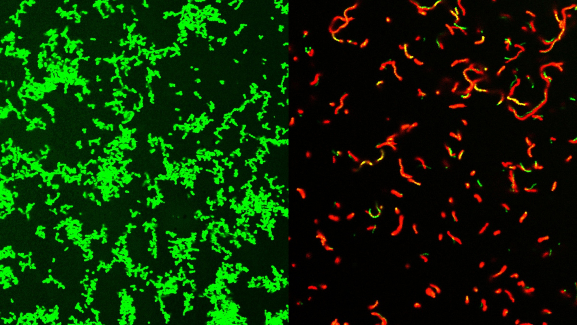
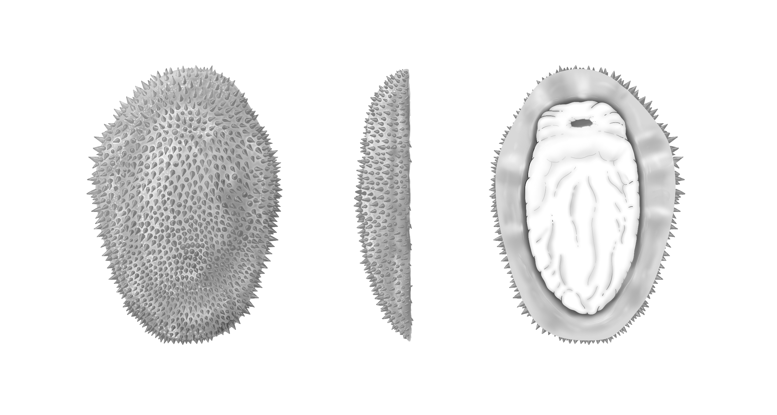
A newly discovered extinct mollusk species that skulked along the ocean floor half a billion years ago is offering new insights into the early days of this diverse group of animals. Fossils from Shishania aculeata indicate that some early mollusks were flat, armored, slug-like creatures that didn’t have the signature shells we see on today’s snails and bivalves. This species was also covered with hollow cone-shaped spines called sclerites. The findings are detailed in a study published August 1 in the journal Science.
Shishania was discovered thanks to some well-preserved fossils uncovered in the Yunnan Province in southern China. The newly named species dates back to the early Cambrian Period–roughly 514 million years ago. The specimens of Shishania that the team studied are a few centimeters long and the spiky cones are made of chitin. This crunchy material is also found in the shells of modern insects, crabs, and even some mushrooms.
The fossils that were preserved upside down, indicates that it likely had a muscular foot similar to a slug. Shishania would have used that leg to creep around the seafloor. Unlike most mollusks, it lacked a shell that covered its body.
Living mollusks come in a wide array of forms–snails, clams, and highly intelligent cephalopods like squids and octopuses. All of this biodiversity developed very quickly during the Cambrian Explosion. This event about 530 million years ago was when all of the major groups of animals were rapidly diversifying. However, due to this accelerated pace of change, few fossils have been left behind to tell the story of early mollusk evolution. The team believes that Shishania represents a very early stage in molluscan evolution.
“Trying to unravel what the common ancestor of animals as different as a squid and oyster looked like is a major challenge for evolutionary biologists and paleontologists–one that can’t be solved by studying only species alive today,” study co-author and University of Oxford in England paleontologist Luke Parry said in a statement. “Shishania gives us a unique view into a time in mollusc evolution for which we have very few fossils, informing us that the very earliest mollusc ancestors were armored spiny slugs, prior to the evolution of the shells that we see in modern snails and clams.”
Shishania’s body was made of soft tissues that typically don’t preserve well in the fossil record. This made the specimens a bit challenging to study, since several were poorly preserved.
“At first I thought that the fossils, which were only about the size of my thumb, were not noticeable, but I saw under a magnifying glass that they seemed strange, spiny, and completely different from any other fossils that I had seen,” Guangxu Zhang, a study co-author and recent PhD graduate from Yunnan University in China who discovered the fossils, said in a statement. “I called it ‘the plastic bag’ initially because it looks like a rotting little plastic bag. When I found more of these fossils and analyzed them in the lab I realized that it was a mollusc.”
Complete specimen of Shishania aculeata seen from the dorsal (top) side (left). Spines covering the body of Shishania aculeata (right). CREDIT: G Zhang/L Parry.
Shishania’s spines show an internal system of canals that are less than one hundredth of a millimeter in diameter. The cones were secreted at their base by microvilli–tiny protrusions of cells that increase surface area. Microvilli are found on the human tongue and in the intestines where they help the body absorb food.
“We found microscopic details inside the conical spines covering the body of Shishania that show how they were secreted in life,” said Parry. “This sort of information is incredibly rare, even in exceptionally preserved fossils.”
The team likens Shishania’s method of secreting hard parts to a natural 3D printer that can change its body parts depending on what the animal needs. This method allows several invertebrates to secrete hard parts that do everything from providing defense to helping it scoot around.
Chitons–the hard spines and bristles in some modern mollusks–are made of the mineral calcium carbonate instead of the organic chitin that is in Shishania. Similar chitinous bristles can be found in some more obscure groups of animals including brachiopods and bryozoans. These animals along with mollusks and annelids (modern earthworms and their relatives) form the group Lophotrochozoa. “Shishania tells us that the spines and spicules we see in chitons and aplacophoran mollusks today actually evolved from organic sclerites like those of annelids,” said Parry. “These animals are very different from one another today and so fossils like Shishania tell us what they looked like deep in the past, soon after they had diverged from common ancestors.”
While humans won’t be regenerating entire limbs like sea stars, some new genetic work with fruit flies has yielded some surprising results. A team from the University of Tokyo found that certain genes from simple organisms that help them regenerate body parts and tissues can be transferred into other animals. These genes then suppressed an intestinal issue in the flies and could potentially reveal some new mechanisms for rejuvenation in more complex organisms. The findings are detailed in a stud
While humans won’t be regenerating entire limbs like sea stars, some new genetic work with fruit flies has yielded some surprising results. A team from the University of Tokyo found that certain genes from simple organisms that help them regenerate body parts and tissues can be transferred into other animals. These genes then suppressed an intestinal issue in the flies and could potentially reveal some new mechanisms for rejuvenation in more complex organisms. The findings are detailed in a study published August 1 in the journal BMC Biology.
Some animals including jellyfish and flatworms can regenerate their whole bodies. While scientists still don’t really know how, there are possibly specific genes that allow regeneration. These same genes may also maintain long-term stem cell functions.
Stem cells can divide and renew themselves over a long period of time and are kind of like a skeleton key. While they aren’t necessarily specialized, they can potentially become more specialized cells, including blood cells and brain cells, over time. Mammals and insects who have very limited regenerative skills may have lost these genes over the course of evolution.
“It is unclear whether reintroducing these regeneration-associated genes in low regenerative animals could affect their regeneration and aging processes,” study co-author and University of Tokyo Graduate School of Pharmaceutical Sciences biologist Yuichiro Nakajima said in a statement.
In this new study, Nakajima and the team focused on the group of genes that is unique to animals with high regenerative capacity like flatworms. These genes are called HRJDs, or highly regenerative species-specific JmjC domain-encoding genes. They transferred the HRJDs into the fruit fly (Drosophila melanogaster) and tracked their health with a blue dye. They nicknamed the fly Smurf, thanks to this hue.
Initially, they hoped that these HRJD-boosted fruit flies would regenerate tissue if injured. This didn’t happen. However, the team had a fruit fly intestine expert Hiroki Nagai onboard, who noticed something else. There were some novel phenotypes–or the characteristics like eye color or hair color that comes from a specific gene.
“HRJDs promoted greater intestinal stem cell division, whilst also suppressing intestinal cells that were mis-differentiating, or going wrong in aged flies,” said Nakajima.
This is different to how antibiotics may suppress the mis-differentiated intestinal cells, but suppress intestinal stem cell division.
“For this reason, HRJDs had a measurable effect on the lifespans of fruit flies, which opens the door, or at least provides clues, for the development of new anti-aging strategies,” said Nakajima. “After all, human and insect intestines have surprisingly much in common on a cellular level.”
Fruit flies are famous test subjects in biological research. They share 75 percent of the genes that cause diseases in humans, reproduce quickly, and their genetic code is fairly easy to change. However, even with their relatively short lives and rapid-fire reproduction and maturating rates, it still took about two months to study their full aging process.
In future studies, the team would like to take a closer look at how HRJD’s work on a molecular level.
“Details of the molecular workings of HRJDs are still unresolved. And it’s unclear whether they work alone or in combination with some other component,” said Nakajima. “Therefore, this is just the start of the journey, but we know now that our modified fruit flies can serve as a valuable resource to uncover unprecedented mechanisms of stem cell rejuvenation in the future. In humans, intestinal stem cells decrease in activity with age, so this research is a promising avenue for stem cell-based therapies.”