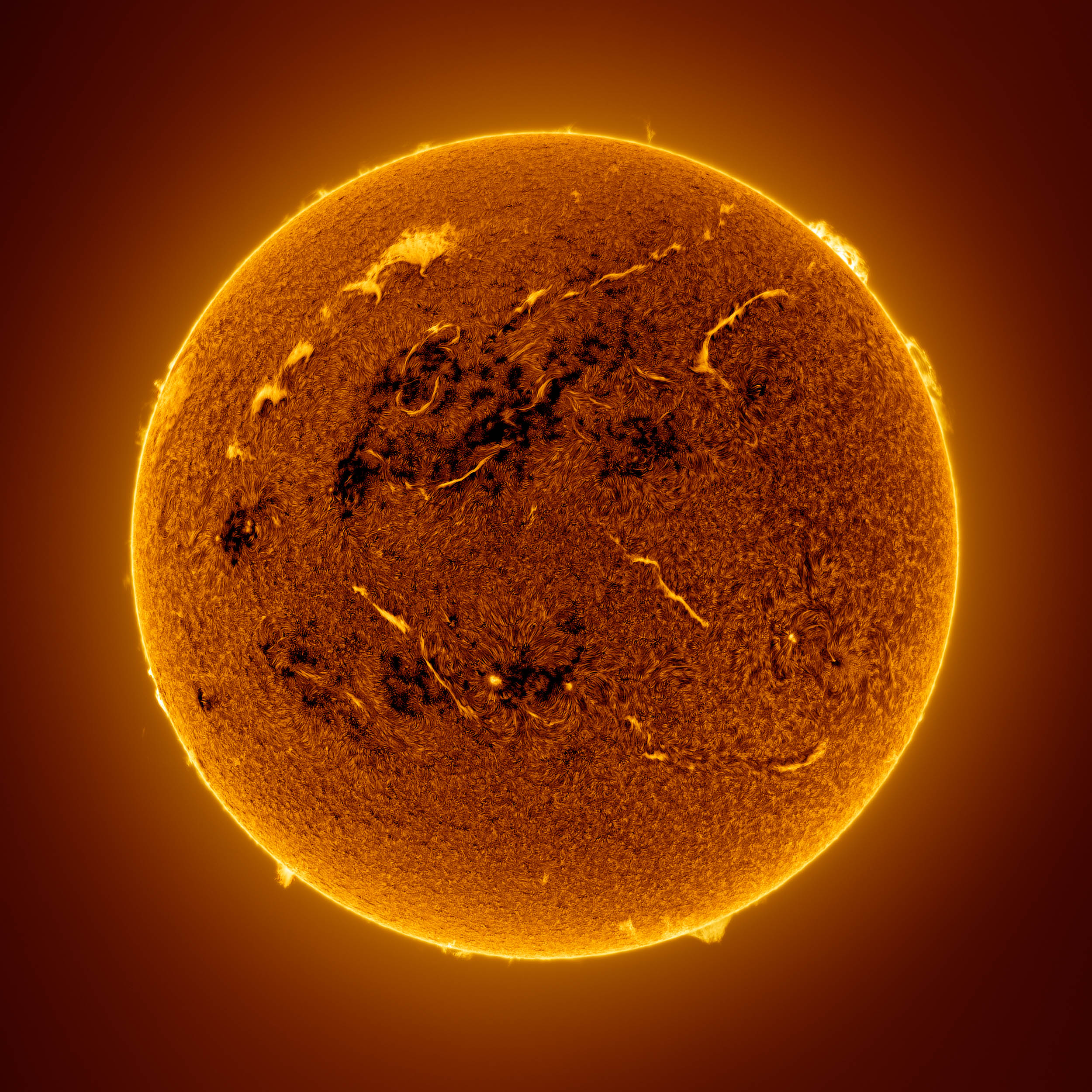
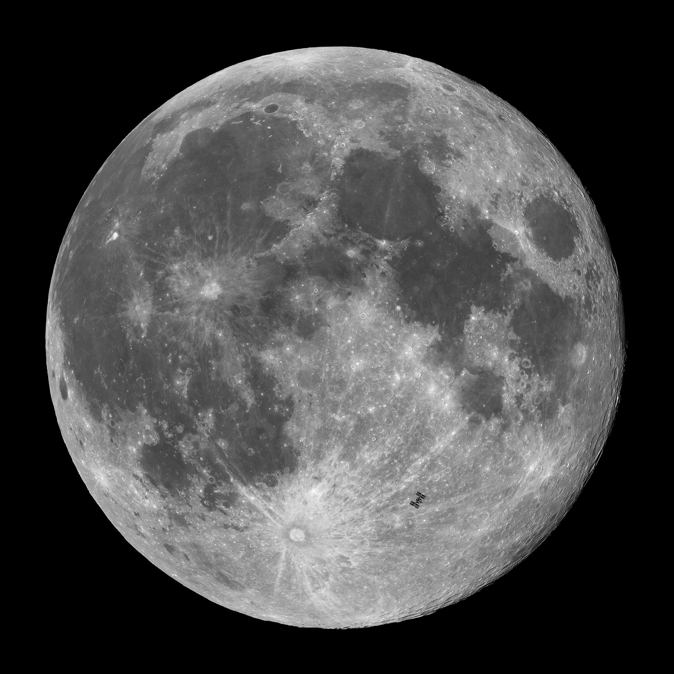
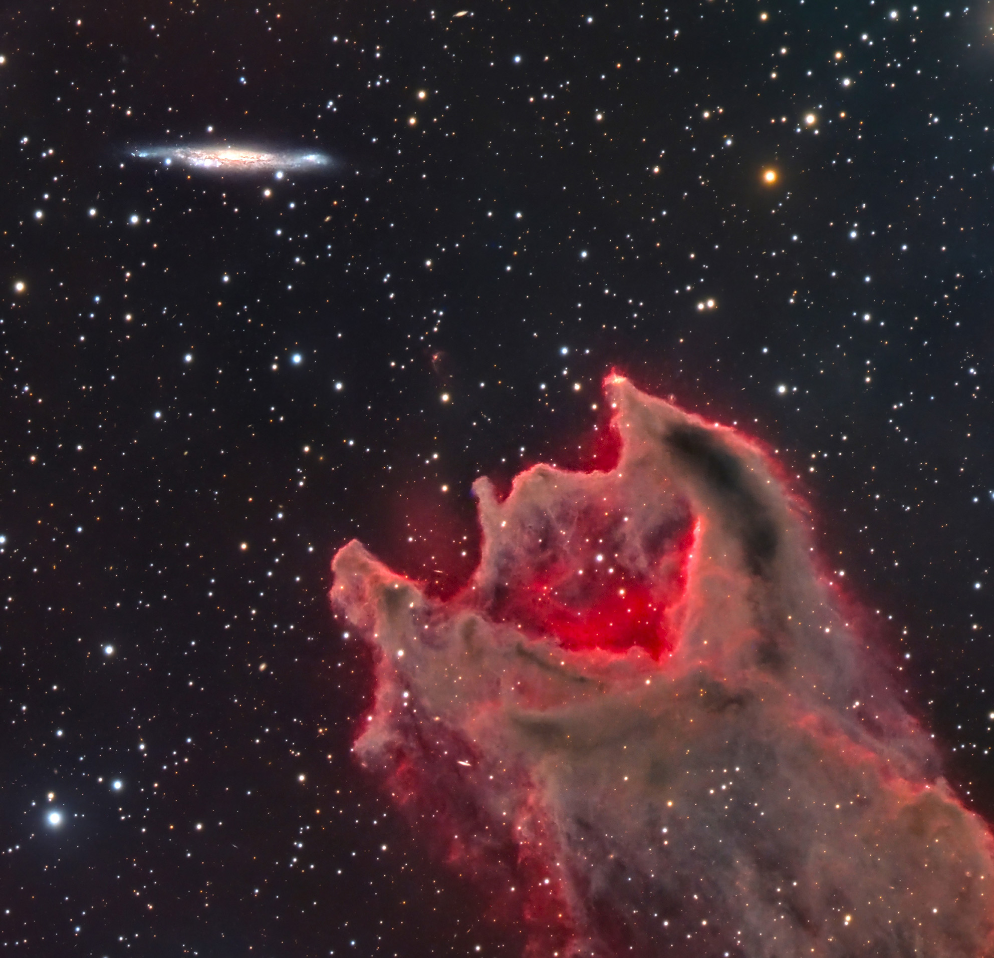
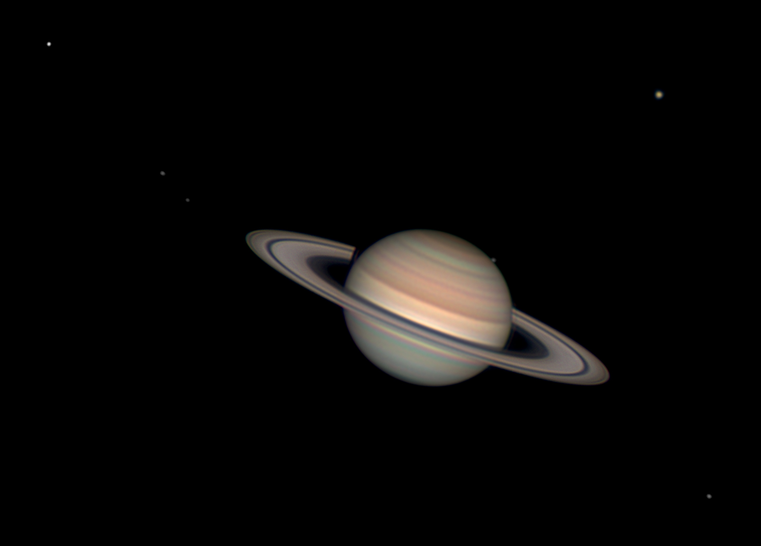
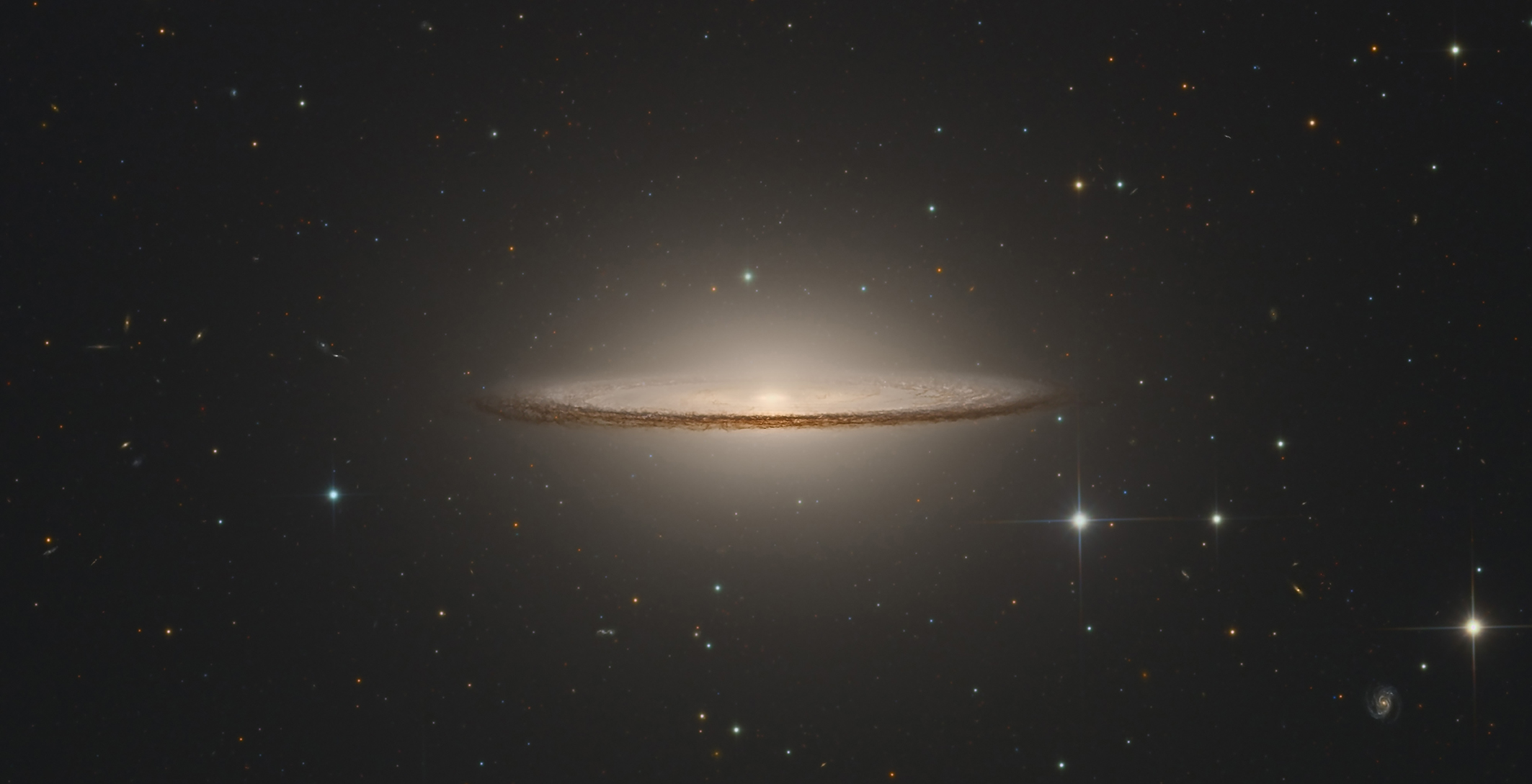
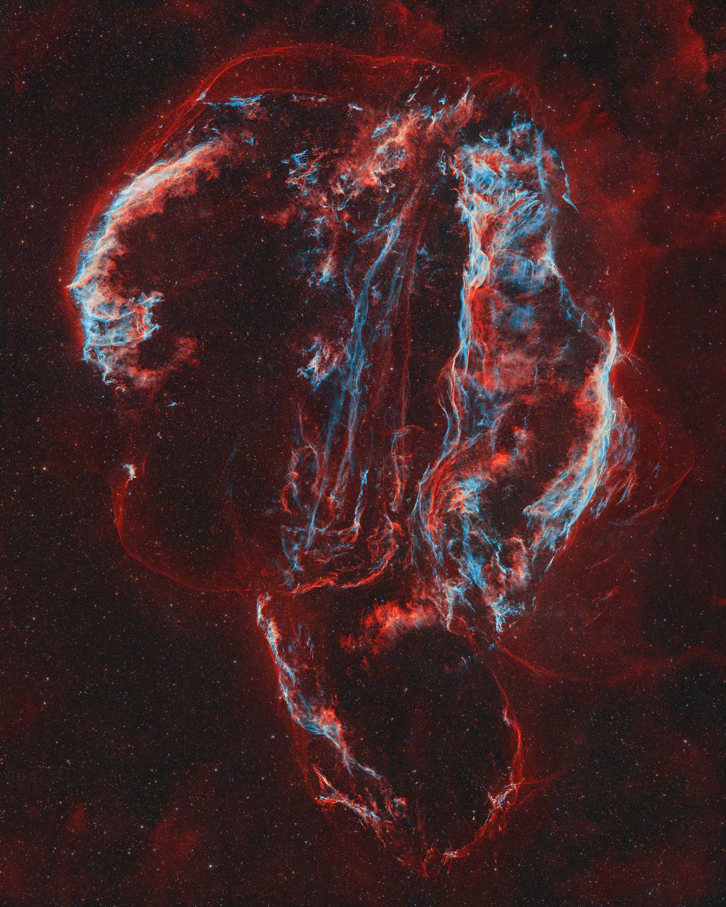
A galactic worm gobbles stars. A plasma whale slides across the sun‘s surface. And an eerie dragon dances with an aurora. It’s not the plot to a fantasy novel, it’s our incredible universe captured in stunning detail.
The Royal Observatory Greenwich has announced the shortlisted images for the 2024 Astronomy Photographer of the Year. The finalists were selected from more than 3,500 images submitted from professional and amateur photographers from 58 countries. The winner will be announced September 12 and an exhibition of the top images will be on display in London at the National Maritime Museum starting September 14.
When you learn about the moon in school, you’re generally taught that its gravity is insufficient to capture and retain any significant atmosphere. The moon is nonetheless surrounded by a thin, ephemeral halo of gasses—an exosphere.
This surprising fact was first discovered using instruments carried by astronauts who visited the moon with the Apollo program. The moon’s weak gravity means that the exosphere’s constituent atoms are constantly draining away into space—and, as such, its continuous presence means that the supply of these atoms is being constantly replenished.
A new study published in Science Advances on August 2 looks at exactly how this replenishment happens. It examines a group of elements whose presence in the lunar atmosphere might come as a surprise to anyone who’s studied chemistry: alkali metals.
Alkali metals form the first group of the periodic table, and include lithium, sodium, potassium, rubidium, and caesium (along with francium, which is never found in macroscopic quantities because it’s so radioactive). Why is their presence a surprise? On Earth, they’re famous for their reactivity, as evidenced by the classic high school demonstration of what a piece of sodium does when it encounters water. On the moon, however, things are very different.
As Prof. Nicole Nie, lead author of the paper, tells Popular Science, “In lunar soils and rocks, alkali metals are bound in minerals, forming stable chemical bonds with oxygen and other elements. But when they are released from the surface, they usually become neutral atoms. There is no liquid water or substantial atmosphere [on the moon], so these metals can remain in their elemental form—[and] because the number of atoms in the lunar atmosphere is so small, the atoms can travel a long distance freely without colliding with one another.”
This does, however, raise the question of how the atoms are released from the surface in the first place. The paper seeks to answer this question—and, specifically, the relative contributions of three processes known collectively as “space weathering.” The uniting factor in these three processes is that they involve something striking the lunar surface and knocking the alkali metal elements out of the mineral compounds in which they’re bound. (These processes also release other elements, but the volatility of alkali metals makes them particularly easy to liberate.)
The first of these processes is micrometeorite impacts, where tiny pieces of space debris rain down with sufficient force to vaporize a small piece of the lunar surface and launch its component atoms into orbit. The second is ion sputtering, where charged particles driven by the solar wind strike the lunar surface. And finally there’s photon-stimulated desorption, where it’s high-energy photons from the sun that knock the alkali metals loose.
As the paper notes, while each process has been well-characterized, previous research has “not conclusively disentangled their [relative] contributions” to the lunar atmosphere. To go about doing this, Nie and her team went right back to the source of the question: the Apollo program. The various crewed missions to the moon in the late 1960s and early ‘70s brought back a total of 382 kg of lunar soil samples, and decades later, these samples are still revealing their secrets to researchers. Nie’s study involved examining 10 samples from five different Apollo missions, including several from Apollo 8, the first manned moon landing.
The team used these samples to look at the relative proportions of different isotopes of potassium and rubidium in the soil. (Sodium and cesium only have one stable isotope each, while lithium is less volatile than its heavier cousins.) As Nie explains to Popular Science, “Lighter isotopes of an element are preferentially released during these processes, leaving the lunar soils with relatively heavier isotopic compositions. For elements that are affected by space weathering, we would expect lunar soils to show heavy isotopic compositions, compared to deeper rocks that are not affected by the process.”
The different space weathering processes produce different ratios of isotopes, and the team’s results indicate that it appears that micrometeorite impacts make the largest contribution to the lunar atmosphere, “likely contributing more than 65% of atmospheric [potassium] atoms, with ion sputtering accounting for the rest.”
This provides a valuable insight into how the moon’s atmosphere has evolved over billions of years—while its composition may well vary over shorter timescales, these results suggest that in the long run, micrometeorite impacts play the dominant role in the constant replenishment of the atmosphere. The study also points to how similar research might be carried out on other objects similar to the moon, like Phobos, one of Mars’s two satellites.
For decades, clinicians have been sounding the alarm about pathogens that are increasingly becoming more resistant to drugs currently available. This makes them more dangerous and according to the Centers for Disease Control and Prevention (CDC), over 2.8 million antimicrobial-resistant infections occur in the US every year. More than 35,000 people die from these infections. To combat this, newer antimicrobial compounds will be needed to replace the ones that bacteria have become resistant to.
Molecular microbiologists Scott Hultgren and Michael Caparon from Washington University School of Medicine in St. Louis and chemist Fredrik Almqvist from the University of Umeå in Sweden collaborated on this new family of compounds called GmPcides.
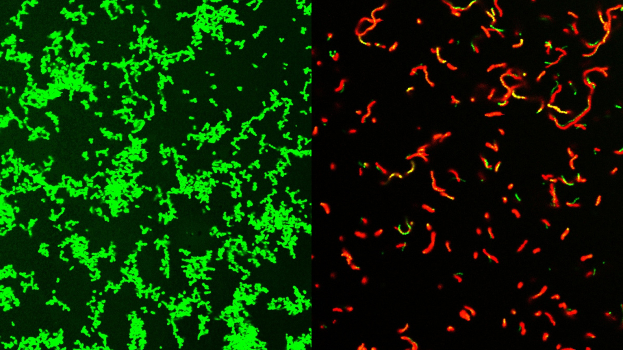
GmPcides work by targeting gram-positive bacteria. These types of bacteria can cause various drug-resistant staph infections, toxic shock syndrome, and other bacterial illnesses that can turn deadly.
“All of the gram-positive bacteria that we’ve tested have been susceptible to that compound. That includes enterococci, staphylococci, streptococci, C. difficile, which are the major pathogenic bacteria types,” Caparon said in a statement. “The compounds have broad-spectrum activity against numerous bacteria.”
The resulting compound also had infection-fighting properties against multiple types of bacteria. Some of their earlier research showed that GmPcides can kill bacteria strains in petri dish experiments.
In this new study, they took those petri dish experiments one step further by testing how compounds work on necrotizing soft-tissue infections. These fast-spreading infections usually involve multiple types of gram-positive bacteria. Necrotizing fasciitis–or flesh-eating disease–is the best known of these infections. It can rapidly damage tissue so severely that limb amputation is often necessary to control its spread. Roughly 20 percent of patients with flesh-eating disease die.
The team focused on one pathogen that is responsible for about 500,000 deaths every year–Streptococcus pyogenes. A group of mice was infected with S. pyogenes. One group was treated with GmPcide, while the other wasn’t. Those that received the GmPcide treatment fared better than the untreated mice in almost every metric. They lost less weight, had smaller ulcers, and fought off the infection faster. Damaged areas of skin also appeared to heal quicker post-infection.
While it is still not fully clear how GmPcides did all of this, a microscopic examination showed that the treatment has a significant effect on bacterial cell membranes. These are the outer wrapping of the microbes.
“One of the jobs of a membrane is to exclude material from the outside,” Caparon said. “We know that within five to ten minutes of treatment with GmPcide, the membranes start to become permeable and allow things that normally should be excluded to enter into the bacteria, which suggests that those membranes have been damaged.”
This can alter the bacteria’s own functions, including actions that damage the host and make the bacteria less effective at taking down the host’s immune response to infections.
GmPcides also may be less likely to lead to drug-resistant strains. The experiments designed to create resistant bacteria found that very few cells can withstand treatment. This means they are less likely to pass on their advantages to the next generation of bacteria.
The road ahead
According to Caparon, there are still numerous steps before GmPcides will be available at your local pharmacy. The team has patented the compound used and licensed it to QureTech Bio, a company that Caparon, Hultgren and Almqvist have an ownership stake in. The license was contingent on the expectation that they will collaborate with a separate company that can manage the pharmaceutical development and clinical trials to bring it to market.
According to the team, the kind of collaborative science that created GmPcides will be needed to treat the problems like antimicrobial resistance. “Bacterial infections of every type are an important health problem, and they are increasingly becoming multi-drug resistant and thus harder to treat,” Hultgren said in a statement. “Interdisciplinary science facilitates the integration of different fields of study that can lead to synergistic new ideas that have the potential to help patients.”
Penguins can’t fly. And while their wings may seem to be purely decorative, these appendages actually play a larger role in their evolutionary history. A fossil penguin species named Pakudyptes hakataramea bridges a gap between penguins that have gone extinct and those living today. Some of its bones show how these wings evolved to help penguins become such speedy swimmers. The findings are described in a study published July 31 in the Journal of the Royal Society of New Zealand.
Pakudyptes lived in present-day New Zealand’s South Island about 24 million years ago. It was very small, roughly the same size as the little blue penguin–or kororā living today. At only 9.8 inches tall and 2.2 pounds, Pakudyptes are among the smallest known penguin species to ever live on Earth.
Interestingly, Pakudyptes did have the physical adaptations that allowed them to dive into the water, despite being such an early penguin species. In the study, a team of scientists from the University of Otago in New Zealand, and Japan’s Ashoro Museum of Paleontology, Okayama University of Science, and Osaka University examined three bones. The humerus, femur, and ulna were discovered during several field trips in 1987 by the late paleontologist Ewan Fordyce in the Hakataramea Valley, in the Canterbury region of the South Island.
They found that Pakudyptes fills in a morphological gap between modern and fossil penguins who are now extinct.
“In particular, the shape of the wing bones differed greatly, and the process by which penguin wings came to have their present form and function remained unclear,” study co-author and Ashoro Museum of Paleontology paleontologist Tatsuro Ando said in a statement.
The humerus and ulna bones show how the penguins’ wings have evolved.
“Surprisingly, while the shoulder joints of the wing of Pakudyptes were very close to the condition of the present-day penguin, the elbow joints were very similar to those of older types of fossil penguins,” said Ando. “Pakudyptes is the first fossil penguin ever found with this combination, and it is the ‘key’ fossil to unlocking the evolution of penguin wings.”
Otago’s Faculty of Dentistry analyzed the fossil’s internal bone structure alongside data on living penguins from the Okayama University of Science. They found that Pakudyptes had microanatomical features that suggest they could dive. Modern penguins are well known for their excellent swimming abilities. Their bullet-like swimming skills are largely due to the dense, thick bones that add to their buoyancy during diving.
In Pakudyptes, the bone cortex was reasonably thick. However, the medullary cavity–which contains bone marrow–was open. This is similar to the living little blue penguin, which usually swims in shallow waters.
Pakudyptes’ diving and swimming likely comes down to the distinctive combination of its bones. The humerus and ulna show spots for attachment of muscles and ligaments, which reveal how the wings were used to swim and move underwater.
“Penguins evolved rapidly from the Late Oligocene to Early Miocene and Pakudyptes is an important fossil from this period,” study co-author Carolina Loch from Otago’s Faculty of Dentistry said in a statement. “Its small size and unique combination of bones may have contributed to the ecological diversity of modern penguins.”
New analysis of a 3,500-year-old mummy known as the “Screaming Woman” may revise what makes for a “good” versus “bad” mummification—and potentially solve a mystery that has perplexed Egyptologists for years.
During the 21st and 22nd Egyptian dynasties, priests oversaw the relocation of a trove of dynastic remains to the Deir el-Bahari Royal Cache in Thebes (near modern-day Luxor). While initial excavations began in 1881, the Metropolitan Museum of New York conducted a follow-up investigation into adjacent crypts in 1935. It was then that archeologists first uncovered the tomb of Senmut, the royal architect and rumored lover of Queen Hatshepsut (1479-1458 BCE). But beneath Senmut’s resting place lay another chamber, this one containing his mother, Hat-Nufer, and multiple unidentified relatives.
Recent examinations into one of those wooden coffins revealed a striking figure—the mummy of an older (for the time), richly adorned woman with a mouth frozen open as if screaming. Although not the first mummy found with such an expression, the anonymous woman’s anatomy and the preservation techniques used on her raised a question for experts. Usually, an open mouth is evidence of a poorly performed mummification, but this didn’t make sense given the Screaming Woman’s royal interment. Now, Egyptologists may finally have at least some answers about the Screaming Woman.
Radiology professor Sahar Saleem and colleagues at Cairo University’s Kasr Al Ainy Hospital have published a new study in the journal Frontiers in Medicine that provides never-before-seen, detailed looks at the mummy along with reliable theories about her health near the end of her life. Thanks to CT imaging, infrared spectroscopy, scanning electron microscopy, and other equipment, Saheer’s team theorize the open mouth may not be due to a shoddy burial, but rather the result of a cadaveric spasm in her final moments.
Scans indicate the woman, approximately 48-years-old, lacked multiple teeth at her time of death—these, however, were lost earlier in her life due to evidence of bone resorption, which occurs when an empty tooth socket reheals. According to Saleem, these may have even been extracted by a professional, as dentistry originated in ancient Egypt. Evidence of bone spurs on her vertebrae also indicates mild spine arthritis.
When it comes to the Screaming Woman’s bodily mummification, a surprising detail sticks out from everything else—the lack of an embalming incision. Egyptologists have long believed classic New Kingdom (1550-1069 BCE) mummifications entailed the removal of a cadaver’s organs except their heart, but the Screaming Woman appeared to still possess them when she was buried. Because of this, Saleen theorizes leaving organs inside a body may actually have been sometimes customary at the time.
As for how she was prepared for mummification, Fourier transform infrared spectroscopy (FTIR) scans of the mummy’s skin revealed the presence of juniper and frankincense, luxuries that Egyptians would need to import from Southern Arabia, East Africa, or the Eastern Mediterranean. The woman’s natural hair was dyed with henna and juniper, but she also wore a long wig for the afterlife made from date palm fingers treated with albite crystals, magnetite, and quartz. These were often used to stiffen the wig’s locks and make them appear black to indicate a more youthful appearance.
“These findings support the ancient trade of embalming materials in ancient Egypt,” Saleem said in an accompanying statement, noting a previous expedition led by Queen Hatshepsut brought back frankincense, while Tutankhamun’s tomb also contained frankincense and juniper.
These embalming methods, combined with her well-preserved appearance, “contradicts the traditional belief that a failure to remove her inner organs implied poor mummification,” said Saleem.
While a definitive answer about the Screaming Woman’s cause of death remains a mystery, Saleem’s work indicates practices like organ removal weren’t always a defining feature of professional mummifications. If nothing else, the spooky visage is likely not due to a bad mummification job. Regardless, Saleem calls the Screaming Woman a “true ‘time capsule’ of the way that she died and was mummified.”
CORRECTION 8/2/2024 6:47AM: A previous version of this article incorrectly dated the Screaming Woman mummy as 2,500-years-old.
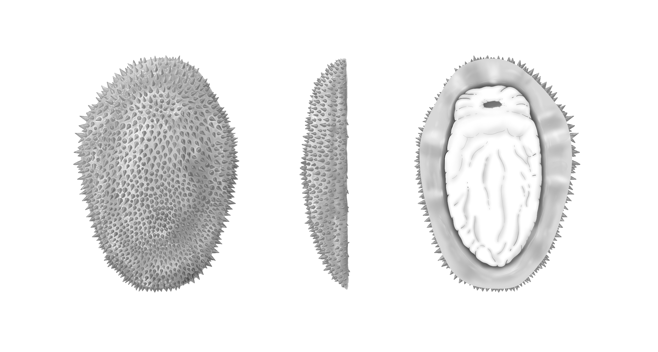
A newly discovered extinct mollusk species that skulked along the ocean floor half a billion years ago is offering new insights into the early days of this diverse group of animals. Fossils from Shishania aculeata indicate that some early mollusks were flat, armored, slug-like creatures that didn’t have the signature shells we see on today’s snails and bivalves. This species was also covered with hollow cone-shaped spines called sclerites. The findings are detailed in a study published August 1 in the journal Science.
Shishania was discovered thanks to some well-preserved fossils uncovered in the Yunnan Province in southern China. The newly named species dates back to the early Cambrian Period–roughly 514 million years ago. The specimens of Shishania that the team studied are a few centimeters long and the spiky cones are made of chitin. This crunchy material is also found in the shells of modern insects, crabs, and even some mushrooms.
The fossils that were preserved upside down, indicates that it likely had a muscular foot similar to a slug. Shishania would have used that leg to creep around the seafloor. Unlike most mollusks, it lacked a shell that covered its body.
Living mollusks come in a wide array of forms–snails, clams, and highly intelligent cephalopods like squids and octopuses. All of this biodiversity developed very quickly during the Cambrian Explosion. This event about 530 million years ago was when all of the major groups of animals were rapidly diversifying. However, due to this accelerated pace of change, few fossils have been left behind to tell the story of early mollusk evolution. The team believes that Shishania represents a very early stage in molluscan evolution.
“Trying to unravel what the common ancestor of animals as different as a squid and oyster looked like is a major challenge for evolutionary biologists and paleontologists–one that can’t be solved by studying only species alive today,” study co-author and University of Oxford in England paleontologist Luke Parry said in a statement. “Shishania gives us a unique view into a time in mollusc evolution for which we have very few fossils, informing us that the very earliest mollusc ancestors were armored spiny slugs, prior to the evolution of the shells that we see in modern snails and clams.”
Shishania’s body was made of soft tissues that typically don’t preserve well in the fossil record. This made the specimens a bit challenging to study, since several were poorly preserved.
“At first I thought that the fossils, which were only about the size of my thumb, were not noticeable, but I saw under a magnifying glass that they seemed strange, spiny, and completely different from any other fossils that I had seen,” Guangxu Zhang, a study co-author and recent PhD graduate from Yunnan University in China who discovered the fossils, said in a statement. “I called it ‘the plastic bag’ initially because it looks like a rotting little plastic bag. When I found more of these fossils and analyzed them in the lab I realized that it was a mollusc.”
Shishania’s spines show an internal system of canals that are less than one hundredth of a millimeter in diameter. The cones were secreted at their base by microvilli–tiny protrusions of cells that increase surface area. Microvilli are found on the human tongue and in the intestines where they help the body absorb food.
“We found microscopic details inside the conical spines covering the body of Shishania that show how they were secreted in life,” said Parry. “This sort of information is incredibly rare, even in exceptionally preserved fossils.”
The team likens Shishania’s method of secreting hard parts to a natural 3D printer that can change its body parts depending on what the animal needs. This method allows several invertebrates to secrete hard parts that do everything from providing defense to helping it scoot around.
Chitons–the hard spines and bristles in some modern mollusks–are made of the mineral calcium carbonate instead of the organic chitin that is in Shishania. Similar chitinous bristles can be found in some more obscure groups of animals including brachiopods and bryozoans. These animals along with mollusks and annelids (modern earthworms and their relatives) form the group Lophotrochozoa. “Shishania tells us that the spines and spicules we see in chitons and aplacophoran mollusks today actually evolved from organic sclerites like those of annelids,” said Parry. “These animals are very different from one another today and so fossils like Shishania tell us what they looked like deep in the past, soon after they had diverged from common ancestors.”
If you have a big pile of Lego blocks, there are a multitude of possibilities for what you can make. If you have just a handful of Legos, you can maybe assemble one creation before you run out. But as your pile keeps shrinking, at a certain point you won’t have enough Legos to make anything interesting—just a brick or two to stack together.
Stars and their planets form from large clouds of gas and dust floating in space. First, the cloud’s core collapses under gravity to form the central star, and then the remaining material coalesces into planets in orbit around that center. The key takeaway here is that stars and their planets form from the same stuff—that is, whatever mixture of elements we see in the star tells us about the building blocks available for planets in that system.
The amount of metals (which in astronomer lingo means anything heavier than hydrogen and helium) in a star is known as its metallicity. Stellar metallicity is “one of the first knobs we turn when doing all sorts of simulations of stars, disks, and planets,” says Jonathan Brande, an astronomer at the University of Kansas not involved in the new study.
Metallicity can also tell us how old a star is in the context of the universe’s lifetime. Heavier elements are formed in the cores of stars and the catastrophic supernova explosions of the biggest stars, so it simply takes time to make these materials. Astronomers therefore expect that the first generations of stars had low metallicities, simply because “fewer heavier elements had been formed when they were born,” adds Brande.
As we can see in our solar system, planets are largely made up of elements other than hydrogen and helium. Accordingly, astronomers have long theorized that a star with lower metallicity would have fewer planets, owing to the lack of elemental building blocks to create them. Observations have also shown that Jupiter-like planets “have a strong correlation with metallicity, meaning that the lower the metallicity the less likely you are to form them,” explains lead author Kiersten Boley, an astronomer at The Ohio State University.
The trend of fewer planets for lower metallicity couldn’t continue forever, though. At some point, you’d simply run out of cosmic Lego blocks to make any planet-sized objects. But no one had actually seen evidence of this “metallicity cliff,” the point where stars run out of planetary construction materials—until now.
Following the logic of “lower metallicity means fewer planets,” previous exoplanet-hunting telescopes like Kepler specifically targeted Sun-like stars. This strategy aimed to increase the chances of discovering new planets. As a result, many of the known exoplanets orbit stars with very similar amounts of metals to our Sun.
NASA’s newer exoplanet-specific satellite (and Kepler’s successor), TESS, followed a different plan. “TESS was a game changer,” says Boley. This mission observed stars of all types across the night sky, showing us what exoplanets were like in different parts of the galaxy, around different types of stars, and more.
“Kiersten searched the 100,000 most metal-poor stars with TESS for small planets,” explained Jessie Christiansen, Chief Scientist of NASA’s Exoplanet Science Institute and a co-author on the study, on X. “If the metallicity trends of Kepler and K2 continued past stars with ~1/3 of the heavy elements of the Sun, she should have found 68 super-Earths.”
“Sixty-eight,” emphasized Christiansen. “She found zero.”
Clearly, there is a limit where the building blocks simply run out. “Like other planet types, super-Earth formation is also difficult for metal-poor stars,” says Boley.
Given that the first stars were nearly devoid of metals, it’s possible that those stars simply didn’t have any planets. Planets “likely only began to form around 7 billion years, nearly half the lifetime of the galaxy,” adds Boley.
This has fascinating implications for how long life might have been around in our Milky Way. “We know life arose on earth within a billion years of its formation about 3-4 billion years ago,” says Brande. Based on the new information from TESS, that means life on Earth formed “maybe about as early as it ever could’ve formed given galactic conditions,” he adds.
While humans won’t be regenerating entire limbs like sea stars, some new genetic work with fruit flies has yielded some surprising results. A team from the University of Tokyo found that certain genes from simple organisms that help them regenerate body parts and tissues can be transferred into other animals. These genes then suppressed an intestinal issue in the flies and could potentially reveal some new mechanisms for rejuvenation in more complex organisms. The findings are detailed in a study published August 1 in the journal BMC Biology.
Some animals including jellyfish and flatworms can regenerate their whole bodies. While scientists still don’t really know how, there are possibly specific genes that allow regeneration. These same genes may also maintain long-term stem cell functions.
Stem cells can divide and renew themselves over a long period of time and are kind of like a skeleton key. While they aren’t necessarily specialized, they can potentially become more specialized cells, including blood cells and brain cells, over time. Mammals and insects who have very limited regenerative skills may have lost these genes over the course of evolution.
“It is unclear whether reintroducing these regeneration-associated genes in low regenerative animals could affect their regeneration and aging processes,” study co-author and University of Tokyo Graduate School of Pharmaceutical Sciences biologist Yuichiro Nakajima said in a statement.
In this new study, Nakajima and the team focused on the group of genes that is unique to animals with high regenerative capacity like flatworms. These genes are called HRJDs, or highly regenerative species-specific JmjC domain-encoding genes. They transferred the HRJDs into the fruit fly (Drosophila melanogaster) and tracked their health with a blue dye. They nicknamed the fly Smurf, thanks to this hue.
Initially, they hoped that these HRJD-boosted fruit flies would regenerate tissue if injured. This didn’t happen. However, the team had a fruit fly intestine expert Hiroki Nagai onboard, who noticed something else. There were some novel phenotypes–or the characteristics like eye color or hair color that comes from a specific gene.
“HRJDs promoted greater intestinal stem cell division, whilst also suppressing intestinal cells that were mis-differentiating, or going wrong in aged flies,” said Nakajima.
This is different to how antibiotics may suppress the mis-differentiated intestinal cells, but suppress intestinal stem cell division.
“For this reason, HRJDs had a measurable effect on the lifespans of fruit flies, which opens the door, or at least provides clues, for the development of new anti-aging strategies,” said Nakajima. “After all, human and insect intestines have surprisingly much in common on a cellular level.”
Fruit flies are famous test subjects in biological research. They share 75 percent of the genes that cause diseases in humans, reproduce quickly, and their genetic code is fairly easy to change. However, even with their relatively short lives and rapid-fire reproduction and maturating rates, it still took about two months to study their full aging process.
In future studies, the team would like to take a closer look at how HRJD’s work on a molecular level.
“Details of the molecular workings of HRJDs are still unresolved. And it’s unclear whether they work alone or in combination with some other component,” said Nakajima. “Therefore, this is just the start of the journey, but we know now that our modified fruit flies can serve as a valuable resource to uncover unprecedented mechanisms of stem cell rejuvenation in the future. In humans, intestinal stem cells decrease in activity with age, so this research is a promising avenue for stem cell-based therapies.”