FreshRSS

Normální zobrazení

Jsou dostupné nové články, klikněte pro obnovení stránky.
PředevčíremHlavní kanál

3 Questions: Darrell Irvine on making HIV vaccines more powerful

An MIT research team led by Professor Darrell Irvine has developed a novel kind of vaccine adjuvant: a nanoparticle that can help to stimulate the immune system to generate a stronger response to a vaccine. These nanoparticles contain saponin, a compound derived from the bark of the Chilean soapbark tree, along with a molecule called MPLA, each of which helps to activate the immune system.

The adjuvant has been incorporated into an experimental HIV vaccine that has shown promising results in animal studies, and this month, the first human volunteers will receive the vaccine as part of a phase 1 clinical trial run by the Consortium for HIV/AIDS Vaccine Development at the Scripps Research Institute. MIT News spoke with Irvine about why this project required an interdisciplinary approach, and what may lie ahead.

Q: What are the special features of the new nanoparticle adjuvant that help it create a more powerful immune response to vaccination? 

A: Most vaccines, such as the Covid-19 vaccines, are thought to protect us through B cells making protective antibodies. Development of an HIV vaccine has been made challenging by the fact that the B cells that are capable of evolving to produce protective antibodies — called broadly neutralizing antibodies — are very rare in the average person. Vaccine adjuvants are important in this scenario to ensure that when we immunize with an HIV antigen, these rare B cells become activated and get a chance to participate in the immune response.

We particularly discovered that this new adjuvant, which we call SMNP (short for saponin/MPLA nanoparticles), is particularly good at helping more B cells enter germinal centers, the specialized location in lymph nodes where high affinity antibodies are produced. In animal models, SMNP also has shown unique mechanisms of action: Administering antigens with SMNP leads to better antigen delivery to lymph nodes (through increases in lymph flow) and better capture of the antigen by B cells in lymph nodes.

Q: How did your lab, which generally focuses on bioengineering and materials science, end up working on HIV vaccines? What obstacles did you have to overcome in the development of this adjuvant?

A: About 15 years ago, Bruce Walker approached me about getting involved in the HIV vaccine effort, and recruited me to join the Ragon Institute of MGH, MIT, and Harvard as a member of the steering committee. Through the Ragon Institute, I met colleagues in the Scripps Consortium for HIV/AIDS Vaccine Development (CHAVD), and we realized there was a tremendous opportunity to directly contribute to the HIV vaccine challenge, working in partnership with experts in immunogen design, structural biology, and HIV pathogenesis.

As we carried out study after study of SMNP in preclinical animal models, we realized the adjuvant had really amazing effects for promoting anti-HIV antibody responses, and the CHAVD decided this was worth moving forward to testing in humans. A major challenge was transferring the technology out of the lab to synthesize large amounts of the adjuvant under GMP (good manufacturing process) conditions for a clinical trial. The initial contract manufacturing organization (CMO) hired by the consortium to produce SMNP simply couldn’t get a process to work for scalable manufacturing.

Luckily for us, a chemical engineering graduate student, Ivan Pires, whom I co-advise with Paula Hammond, head of MIT’s Department of Chemical Engineering, had developed expertise in one particular processing technique known as tangential flow filtration during his undergraduate training. Leveraging classic chemical engineering skills in thermodynamics and process design, Ivan stepped in and solved the process issues the CMO was facing, allowing the manufacturing to move forward. This to me is what makes MIT great — the ability of our students and postdocs to step up and solve big problems and make big contributions when the need arises.

Q: What other diseases could this approach be useful for? Are there any plans to test it with other types of vaccines?

A: In principle, SMNP may be helpful for any infectious disease vaccine where strong antibody responses are needed. We are currently sharing the adjuvant with about 30 different labs around the world, who are testing it in vaccines against many other pathogens including Epstein-Barr virus, malaria, and influenza. We are hopeful that if SMNP is safe and effective in humans, this will be an adjuvant that can be broadly used in infectious disease trials.

© Photo: Steve Boxall


An MIT research team led by Professor Darrell Irvine has developed a novel kind of vaccine adjuvant: a nanoparticle that can help to stimulate the immune system to generate a stronger response to a vaccine. An HIV vaccine that includes this adjuvant will be tested in clinical trials this month.

Capsid of HIV-1 behaves like cell’s cargo receptor to enter the nucleus

Retroviruses cannot replicate on their own — they must insert their genetic code into the DNA of a host and exploit the host cell's resources to make more copies of themselves, furthering infection. Some retroviruses only infect cells as they divide, when the nuclear envelope that protects the host's genetic material breaks down, making it easily accessible. HIV-1 is a type of retrovirus, called a lentivirus, that can infect non-dividing cells.

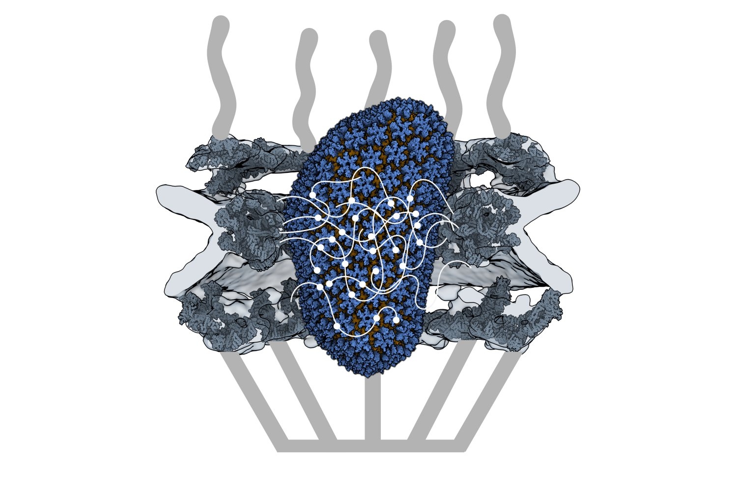
HIV-1 delivers its genome into the nucleus by packaging it into a large, cone-shaped structure called a capsid — but the exact mechanism has remained elusive for decades. Travel through the nuclear envelope occurs through, and is regulated by, nuclear pores, doughnut-shaped protein assemblies. Human cells have about 2,000 nuclear pores perforating the nuclear envelope. Some earlier evidence suggested that the capsid remains intact during its delivery into the nucleus — but this created a dimensional conundrum. The cone-shaped HIV-1 capsid is about 120 nanometers long and 60 nm wide — too large, researchers thought, to fit through the opening of the nuclear pore, measured at only 43 nm wide.

Members of the Schwartz Lab at MIT, in the Department of Biology, became interested in this question when a postdoc in the lab used cryo-electron tomography, slicing up sections of frozen cells to examine structures, to show that nuclear pores in the nuclear envelope are larger than 43nm. They deflate and shrink, it turns out, when removed from their native conditions. In native conditions, the nuclear pore complex is about 60nm wide — wide enough to accommodate the HIV-1 capsid.

Knowing that it could fit, a question remained: How can the capsid navigate the dense mesh of spaghetti-like proteins that act like a sieve in the nuclear pore channel? That spaghetti-like mesh allows small cargo to diffuse through, but prevents large cargo from entering unless it is escorted by proteins called nuclear transport receptors.

In an open-access paper published today in Nature, researchers present evidence that the HIV-1 capsid mimics the cell's transport receptors to traverse the nuclear pore.

To support that conclusion, the researchers showed three things in vitro: that an HIV-1 capsid can deliver cargo through a nuclear pore analog; that the capsid can interact with the sieve of proteins in the nuclear pore channel; and that the capsid targets the nuclear pore in the absence of native transport proteins.

Nuclear transport receptors escort large cargo through the nuclear pore by “batting away” the spaghetti-like mesh of proteins inside the channel — like someone holding your hand and guiding you across a crowded dance floor. The HIV-1 capsid interacts with the spaghetti-like proteins, but its purpose is more like a Trojan horse — the capsid encapsulates the viral cargo, protecting it from detection in the cytoplasm and as it enters the nuclear pore complex.

"What's really amazing about cells is that they are incredibly complex. What's really difficult about studying cells is that they are incredibly complex," jokes co-first author Erika Weiskopf, a graduate student in the Schwartz lab. "Biochemists are constantly trying to find ways to study their system in a simplified context, but still give it a flavor of cell biology."

To do that, the Schwartz lab collaborated with Dirk Görlich, the director of cellular logistics at the Max Planck Institute for Multidisciplinary Sciences. Görlich is a co-senior author on the paper with MIT’s Boris Magasanik Professor of Biology Thomas Schwartz. Görlich's lab has produced concentrated droplets of the spaghetti-like proteins found inside the nuclear pore, and those droplets allow and exclude cargo the same way a nuclear pore will. In experiments, fluorescently-labeled cargo did not enter the droplets, but fluorescently-labeled cargo packaged in an HIV-1 capsid was delivered. This indicated that the capsid could deliver cargo through a nuclear pore. 

Using a biophysical binding assay, the researchers also showed that the HIV-1 capsid interacts with the proteins inside the channel. Different spaghetti-like proteins are found in different channel sections, such as at the cytoplasmic side's entrance or only inside the channel; there are 10 such proteins in human cells. The capsid is a promiscuous binder — it can interact with all the spaghetti-like proteins found in the channel.

The capsid can target the nuclear pore complex even without the cell's transport receptors, indicating that it is not commandeering native transport receptors to find and enter the nuclear pore. The team used a classic assay in the nucleocytoplasmic transport field to collect this evidence: When cells are treated with digitonin, their membranes become porous. Everything in the cytoplasm will leak out of the cells, but the nuclear envelope will remain intact. Despite the absence of native proteins, the capsid was attracted to the nuclear pore complex, a behavior indicative of a nuclear transport receptor.

Although the capsid behaves like a nuclear transport receptor to penetrate the nuclear pore, it is fundamentally different. A transport receptor doesn't need to conceal material for delivery the way the capsid does to avoid detection.

These findings open new lines of inquiry for what the nuclear pore complex is capable of accommodating.

"The HIV-1 capsid is one of the largest things that we now know can go through the nuclear pore complex intact," Weiskopf says. "It raises all kinds of questions — what other things could be going through the pore that we thought was impossible?"

Schwartz said another question is whether all of the 2,000 nuclear pores in human cells are identical or whether there is something that makes certain pores more amenable to allowing the capsid through.

The capsid is also known to be unusually elastic, a property that may be key for passage through the pore. Another interesting question for the field is whether the cone-shaped capsid gains entry into the pore by squeezing through.

Although the team has shown that the capsid can enter the pore, what happens at the other end of the channel is still unknown — whether the capsid fully or partially enters the nucleus or breaks down inside the channel. Weiskopf is working on perturbing parts of the capsid or the spaghetti-like proteins to learn more about which interactions are most important for successful capsid entry.

Although these results have expanded our understanding of the nuclear pore, much remains unknown, both for HIV-1 infection and for the transport process through the nuclear pore complex.  

"The nuclear pore is such an important element of cell biology, we thought it would be interesting to understand it better — and that's how we figured out that the pore is much bigger than we anticipated," Schwartz says. "We will certainly try to see whether we can understand the mechanism of HIV-1 infection, how the capsid is released on the other side of the channel, and what factors are important there — and to what extent you can manipulate it or influence it for therapeutic applications."

This research was carried out, in part, using MIT.nano facilities.

© Image courtesy of the Schwartz Lab.

An HIV-1 capsid penetrates a nuclear pore complex by interacting with the spaghetti-like proteins inside the channel, behaving like the cell’s own cargo transport proteins.

Angela Belcher delivers 2023 Dresselhaus Lecture on evolving organisms for new nanomaterials

“How do we get to making nanomaterials that haven’t been evolved before?” asked Angela Belcher at the 2023 Mildred S. Dresselhaus Lecture at MIT on Nov. 20. “We can use elements that biology has already given us.”

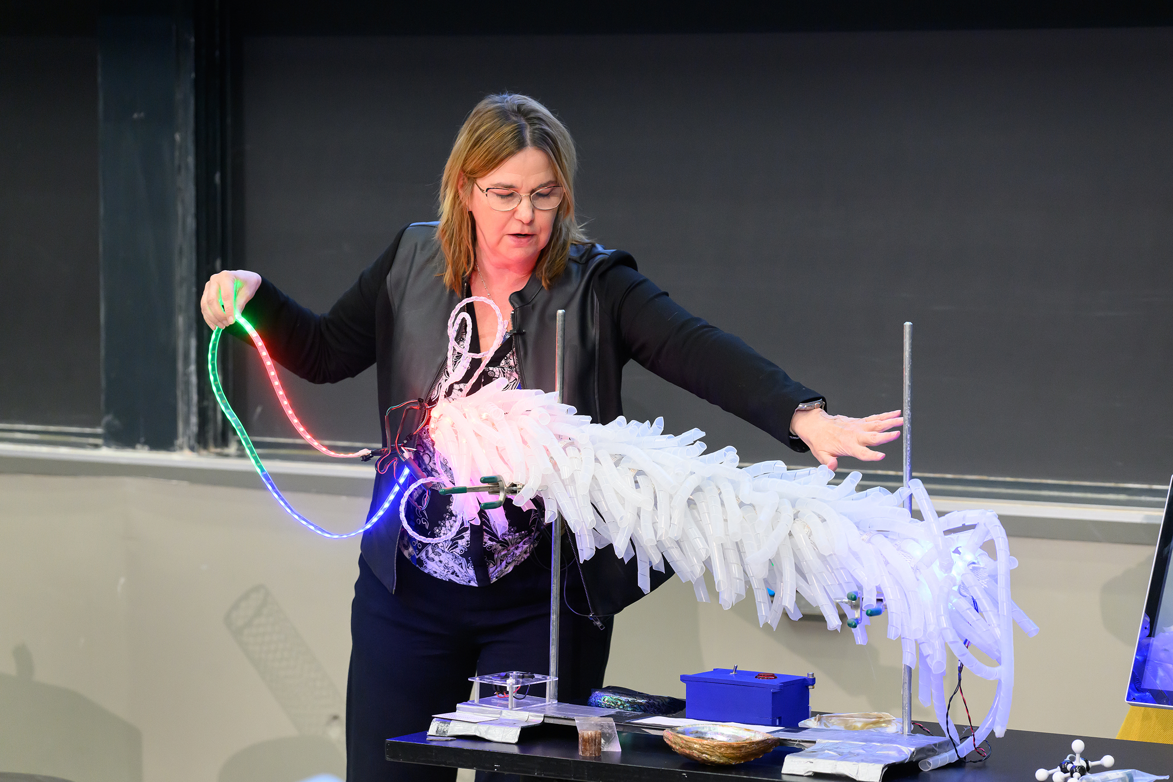
The combined in-person and virtual audience of over 300 was treated to a light-up, 3D model of M13 bacteriophage, a virus that only infects bacteria, complete with a pull-out strand of DNA. Belcher used the feather-boa-like model to show how her research group modifies the M13’s genes to add new DNA and peptide sequences to template inorganic materials.

“I love controlling materials at the nanoscale using biology,” said Belcher, the James Mason Crafts Professor of Biological Engineering, materials science professor, and of the Koch Institute of Integrative Cancer Research at MIT. “We all know if you control materials at the nanoscale and you can start to tune them, then you can have all kinds of different applications.” And the opportunities are indeed vast — from building batteries, fuel cells, and solar cells to carbon sequestration and storage, environmental remediation, catalysis, and medical diagnostics and imaging.

Belcher sprinkled her talk with models and props, lined up on a table at the front of the 10-250 lecture hall, to demonstrate a wide variety of concepts and projects made possible by the intersection of biology and nanotechnology.

Energy storage and environment

“How do you go from a DNA sequence to a functioning battery?” posed Belcher. Grabbing a model of a large carbon nanotube, she explained how her group engineered a phage to pick up carbon nanotubes that would wind all the way around the virus and then fill in with different cathode or anode materials to make nanowires for battery electrodes.

How about using the M13 bacteriophage to improve the environment? Belcher referred to a project by former student Geran Zhang PhD ’19 that proved the virus can be modified for this context, too. He used the phage to template high-surface-area, carbon-based materials that can grab small molecules and break them down, Belcher said, opening a realm of possibilities from cleaning up rivers to developing chemical warfare agents to combating smog.

Belcher’s lab worked with the U.S. Army to produce protective clothing and masks made of these carbon-based virus nanofibers. “We went from five liters in our lab to a thousand liters, then 10,000 liters in the army labs where we’re able to make kilograms of the material,” Belcher said, stressing the importance of being able to test and prototype at scale.

Imaging tools and therapeutics in cancer

In the area of biomedical imaging, Belcher explained, a lot less is known in near-infrared imaging — imaging in wavelengths above 1,000 nanometers — than other imaging techniques, yet with near-infrared scientists can see much deeper inside the body. Belcher’s lab built their own systems to image at these wavelengths. The third generation of this system provides real-time, sub-millimeter optical imaging for guided surgery.

Working with Sangeeta Bhatia, the John J. and Dorothy Wilson Professor of Engineering, Belcher used carbon nanotubes to build imaging tools that find tiny tumors during surgery that doctors otherwise would not be able to see. The tool is actually a virus engineered to carry with it a fluorescent, single-walled carbon nanotube as it seeks out the tumors.

Nearing the end of her talk, Belcher presented a goal: to develop an accessible detection and diagnostic technology for ovarian cancer in five to 10 years.

“We think that we can do it,” Belcher said. She described her students’ work developing a way to scan an entire fallopian tube, as opposed to just one small portion, to find pre-cancer lesions, and talked about the team of MIT faculty, doctors, and researchers working collectively toward this goal.

“Part of the secret of life and the meaning of life is helping other people enjoy the passage of time,” said Belcher in her closing remarks. “I think that we can all do that by working to solve some of the biggest issues on the planet, including helping to diagnose and treat ovarian cancer early so people have more time to spend with their family.”

Honoring Mildred S. Dresselhaus

Belcher was the fifth speaker to deliver the Dresselhaus Lecture, an annual event organized by MIT.nano to honor the late MIT physics and electrical engineering Institute Professor Mildred Dresselhaus. The lecture features a speaker from anywhere in the world whose leadership and impact echo Dresselhaus’s life, accomplishments, and values.

“Millie was and is a huge hero of mine,” said Belcher. “Giving a lecture in Millie’s name is just the greatest honor.”

Belcher dedicated the talk to Dresselhaus, whom she described with an array of accolades — a trailblazer, a genius, an amazing mentor, teacher, and inventor. “Just knowing her was such a privilege,” she said.

Belcher also dedicated her talk to her own grandmother and mother, both of whom passed away from cancer, as well as late MIT professors Susan Lindquist and Angelika Amon, who both died of ovarian cancer.

“I’ve been so fortunate to work with just the most talented and dedicated graduate students, undergraduate students, postdocs, and researchers,” concluded Belcher. “It has been a pure joy to be in partnership with all of you to solve these very daunting problems.”

© Photo: Justin Knight

Professor Angela Belcher pulls a single strand of DNA out of a light-up model of M13 bacteriophage, a virus that only infects bacteria. Belcher’s lab modifies the M13’s genes to add new DNA and peptide sequences to template inorganic materials.

3 Questions: Darrell Irvine on making HIV vaccines more powerful

An MIT research team led by Professor Darrell Irvine has developed a novel kind of vaccine adjuvant: a nanoparticle that can help to stimulate the immune system to generate a stronger response to a vaccine. These nanoparticles contain saponin, a compound derived from the bark of the Chilean soapbark tree, along with a molecule called MPLA, each of which helps to activate the immune system.

The adjuvant has been incorporated into an experimental HIV vaccine that has shown promising results in animal studies, and this month, the first human volunteers will receive the vaccine as part of a phase 1 clinical trial run by the Consortium for HIV/AIDS Vaccine Development at the Scripps Research Institute. MIT News spoke with Irvine about why this project required an interdisciplinary approach, and what may lie ahead.

Q: What are the special features of the new nanoparticle adjuvant that help it create a more powerful immune response to vaccination? 

A: Most vaccines, such as the Covid-19 vaccines, are thought to protect us through B cells making protective antibodies. Development of an HIV vaccine has been made challenging by the fact that the B cells that are capable of evolving to produce protective antibodies — called broadly neutralizing antibodies — are very rare in the average person. Vaccine adjuvants are important in this scenario to ensure that when we immunize with an HIV antigen, these rare B cells become activated and get a chance to participate in the immune response.

We particularly discovered that this new adjuvant, which we call SMNP (short for saponin/MPLA nanoparticles), is particularly good at helping more B cells enter germinal centers, the specialized location in lymph nodes where high affinity antibodies are produced. In animal models, SMNP also has shown unique mechanisms of action: Administering antigens with SMNP leads to better antigen delivery to lymph nodes (through increases in lymph flow) and better capture of the antigen by B cells in lymph nodes.

Q: How did your lab, which generally focuses on bioengineering and materials science, end up working on HIV vaccines? What obstacles did you have to overcome in the development of this adjuvant?

A: About 15 years ago, Bruce Walker approached me about getting involved in the HIV vaccine effort, and recruited me to join the Ragon Institute of MGH, MIT, and Harvard as a member of the steering committee. Through the Ragon Institute, I met colleagues in the Scripps Consortium for HIV/AIDS Vaccine Development (CHAVD), and we realized there was a tremendous opportunity to directly contribute to the HIV vaccine challenge, working in partnership with experts in immunogen design, structural biology, and HIV pathogenesis.

As we carried out study after study of SMNP in preclinical animal models, we realized the adjuvant had really amazing effects for promoting anti-HIV antibody responses, and the CHAVD decided this was worth moving forward to testing in humans. A major challenge was transferring the technology out of the lab to synthesize large amounts of the adjuvant under GMP (good manufacturing process) conditions for a clinical trial. The initial contract manufacturing organization (CMO) hired by the consortium to produce SMNP simply couldn’t get a process to work for scalable manufacturing.

Luckily for us, a chemical engineering graduate student, Ivan Pires, whom I co-advise with Paula Hammond, head of MIT’s Department of Chemical Engineering, had developed expertise in one particular processing technique known as tangential flow filtration during his undergraduate training. Leveraging classic chemical engineering skills in thermodynamics and process design, Ivan stepped in and solved the process issues the CMO was facing, allowing the manufacturing to move forward. This to me is what makes MIT great — the ability of our students and postdocs to step up and solve big problems and make big contributions when the need arises.

Q: What other diseases could this approach be useful for? Are there any plans to test it with other types of vaccines?

A: In principle, SMNP may be helpful for any infectious disease vaccine where strong antibody responses are needed. We are currently sharing the adjuvant with about 30 different labs around the world, who are testing it in vaccines against many other pathogens including Epstein-Barr virus, malaria, and influenza. We are hopeful that if SMNP is safe and effective in humans, this will be an adjuvant that can be broadly used in infectious disease trials.

© Photo: Steve Boxall
An MIT research team led by Professor Darrell Irvine has developed a novel kind of vaccine adjuvant: a nanoparticle that can help to stimulate the immune system to generate a stronger response to a vaccine. An HIV vaccine that includes this adjuvant will be tested in clinical trials this month.

Capsid of HIV-1 behaves like cell’s cargo receptor to enter the nucleus

Retroviruses cannot replicate on their own — they must insert their genetic code into the DNA of a host and exploit the host cell's resources to make more copies of themselves, furthering infection. Some retroviruses only infect cells as they divide, when the nuclear envelope that protects the host's genetic material breaks down, making it easily accessible. HIV-1 is a type of retrovirus, called a lentivirus, that can infect non-dividing cells.

HIV-1 delivers its genome into the nucleus by packaging it into a large, cone-shaped structure called a capsid — but the exact mechanism has remained elusive for decades. Travel through the nuclear envelope occurs through, and is regulated by, nuclear pores, doughnut-shaped protein assemblies. Human cells have about 2,000 nuclear pores perforating the nuclear envelope. Some earlier evidence suggested that the capsid remains intact during its delivery into the nucleus — but this created a dimensional conundrum. The cone-shaped HIV-1 capsid is about 120 nanometers long and 60 nm wide — too large, researchers thought, to fit through the opening of the nuclear pore, measured at only 43 nm wide.

Members of the Schwartz Lab at MIT, in the Department of Biology, became interested in this question when a postdoc in the lab used cryo-electron tomography, slicing up sections of frozen cells to examine structures, to show that nuclear pores in the nuclear envelope are larger than 43nm. They deflate and shrink, it turns out, when removed from their native conditions. In native conditions, the nuclear pore complex is about 60nm wide — wide enough to accommodate the HIV-1 capsid.

Knowing that it could fit, a question remained: How can the capsid navigate the dense mesh of spaghetti-like proteins that act like a sieve in the nuclear pore channel? That spaghetti-like mesh allows small cargo to diffuse through, but prevents large cargo from entering unless it is escorted by proteins called nuclear transport receptors.

In an open-access paper published today in Nature, researchers present evidence that the HIV-1 capsid mimics the cell's transport receptors to traverse the nuclear pore.

To support that conclusion, the researchers showed three things in vitro: that an HIV-1 capsid can deliver cargo through a nuclear pore analog; that the capsid can interact with the sieve of proteins in the nuclear pore channel; and that the capsid targets the nuclear pore in the absence of native transport proteins.

Nuclear transport receptors escort large cargo through the nuclear pore by “batting away” the spaghetti-like mesh of proteins inside the channel — like someone holding your hand and guiding you across a crowded dance floor. The HIV-1 capsid interacts with the spaghetti-like proteins, but its purpose is more like a Trojan horse — the capsid encapsulates the viral cargo, protecting it from detection in the cytoplasm and as it enters the nuclear pore complex.

"What's really amazing about cells is that they are incredibly complex. What's really difficult about studying cells is that they are incredibly complex," jokes co-first author Erika Weiskopf, a graduate student in the Schwartz lab. "Biochemists are constantly trying to find ways to study their system in a simplified context, but still give it a flavor of cell biology."

To do that, the Schwartz lab collaborated with Dirk Görlich, the director of cellular logistics at the Max Planck Institute for Multidisciplinary Sciences. Görlich is a co-senior author on the paper with MIT’s Boris Magasanik Professor of Biology Thomas Schwartz. Görlich's lab has produced concentrated droplets of the spaghetti-like proteins found inside the nuclear pore, and those droplets allow and exclude cargo the same way a nuclear pore will. In experiments, fluorescently-labeled cargo did not enter the droplets, but fluorescently-labeled cargo packaged in an HIV-1 capsid was delivered. This indicated that the capsid could deliver cargo through a nuclear pore. 

Using a biophysical binding assay, the researchers also showed that the HIV-1 capsid interacts with the proteins inside the channel. Different spaghetti-like proteins are found in different channel sections, such as at the cytoplasmic side's entrance or only inside the channel; there are 10 such proteins in human cells. The capsid is a promiscuous binder — it can interact with all the spaghetti-like proteins found in the channel.

The capsid can target the nuclear pore complex even without the cell's transport receptors, indicating that it is not commandeering native transport receptors to find and enter the nuclear pore. The team used a classic assay in the nucleocytoplasmic transport field to collect this evidence: When cells are treated with digitonin, their membranes become porous. Everything in the cytoplasm will leak out of the cells, but the nuclear envelope will remain intact. Despite the absence of native proteins, the capsid was attracted to the nuclear pore complex, a behavior indicative of a nuclear transport receptor.

Although the capsid behaves like a nuclear transport receptor to penetrate the nuclear pore, it is fundamentally different. A transport receptor doesn't need to conceal material for delivery the way the capsid does to avoid detection.

These findings open new lines of inquiry for what the nuclear pore complex is capable of accommodating.

"The HIV-1 capsid is one of the largest things that we now know can go through the nuclear pore complex intact," Weiskopf says. "It raises all kinds of questions — what other things could be going through the pore that we thought was impossible?"

Schwartz said another question is whether all of the 2,000 nuclear pores in human cells are identical or whether there is something that makes certain pores more amenable to allowing the capsid through.

The capsid is also known to be unusually elastic, a property that may be key for passage through the pore. Another interesting question for the field is whether the cone-shaped capsid gains entry into the pore by squeezing through.

Although the team has shown that the capsid can enter the pore, what happens at the other end of the channel is still unknown — whether the capsid fully or partially enters the nucleus or breaks down inside the channel. Weiskopf is working on perturbing parts of the capsid or the spaghetti-like proteins to learn more about which interactions are most important for successful capsid entry.

Although these results have expanded our understanding of the nuclear pore, much remains unknown, both for HIV-1 infection and for the transport process through the nuclear pore complex.  

"The nuclear pore is such an important element of cell biology, we thought it would be interesting to understand it better — and that's how we figured out that the pore is much bigger than we anticipated," Schwartz says. "We will certainly try to see whether we can understand the mechanism of HIV-1 infection, how the capsid is released on the other side of the channel, and what factors are important there — and to what extent you can manipulate it or influence it for therapeutic applications."

This research was carried out, in part, using MIT.nano facilities.

© Image courtesy of the Schwartz Lab.

An HIV-1 capsid penetrates a nuclear pore complex by interacting with the spaghetti-like proteins inside the channel, behaving like the cell’s own cargo transport proteins.

Angela Belcher delivers 2023 Dresselhaus Lecture on evolving organisms for new nanomaterials

“How do we get to making nanomaterials that haven’t been evolved before?” asked Angela Belcher at the 2023 Mildred S. Dresselhaus Lecture at MIT on Nov. 20. “We can use elements that biology has already given us.”

The combined in-person and virtual audience of over 300 was treated to a light-up, 3D model of M13 bacteriophage, a virus that only infects bacteria, complete with a pull-out strand of DNA. Belcher used the feather-boa-like model to show how her research group modifies the M13’s genes to add new DNA and peptide sequences to template inorganic materials.

“I love controlling materials at the nanoscale using biology,” said Belcher, the James Mason Crafts Professor of Biological Engineering, materials science professor, and of the Koch Institute of Integrative Cancer Research at MIT. “We all know if you control materials at the nanoscale and you can start to tune them, then you can have all kinds of different applications.” And the opportunities are indeed vast — from building batteries, fuel cells, and solar cells to carbon sequestration and storage, environmental remediation, catalysis, and medical diagnostics and imaging.

Belcher sprinkled her talk with models and props, lined up on a table at the front of the 10-250 lecture hall, to demonstrate a wide variety of concepts and projects made possible by the intersection of biology and nanotechnology.

Energy storage and environment

“How do you go from a DNA sequence to a functioning battery?” posed Belcher. Grabbing a model of a large carbon nanotube, she explained how her group engineered a phage to pick up carbon nanotubes that would wind all the way around the virus and then fill in with different cathode or anode materials to make nanowires for battery electrodes.

How about using the M13 bacteriophage to improve the environment? Belcher referred to a project by former student Geran Zhang PhD ’19 that proved the virus can be modified for this context, too. He used the phage to template high-surface-area, carbon-based materials that can grab small molecules and break them down, Belcher said, opening a realm of possibilities from cleaning up rivers to developing chemical warfare agents to combating smog.

Belcher’s lab worked with the U.S. Army to produce protective clothing and masks made of these carbon-based virus nanofibers. “We went from five liters in our lab to a thousand liters, then 10,000 liters in the army labs where we’re able to make kilograms of the material,” Belcher said, stressing the importance of being able to test and prototype at scale.

Imaging tools and therapeutics in cancer

In the area of biomedical imaging, Belcher explained, a lot less is known in near-infrared imaging — imaging in wavelengths above 1,000 nanometers — than other imaging techniques, yet with near-infrared scientists can see much deeper inside the body. Belcher’s lab built their own systems to image at these wavelengths. The third generation of this system provides real-time, sub-millimeter optical imaging for guided surgery.

Working with Sangeeta Bhatia, the John J. and Dorothy Wilson Professor of Engineering, Belcher used carbon nanotubes to build imaging tools that find tiny tumors during surgery that doctors otherwise would not be able to see. The tool is actually a virus engineered to carry with it a fluorescent, single-walled carbon nanotube as it seeks out the tumors.

Nearing the end of her talk, Belcher presented a goal: to develop an accessible detection and diagnostic technology for ovarian cancer in five to 10 years.

“We think that we can do it,” Belcher said. She described her students’ work developing a way to scan an entire fallopian tube, as opposed to just one small portion, to find pre-cancer lesions, and talked about the team of MIT faculty, doctors, and researchers working collectively toward this goal.

“Part of the secret of life and the meaning of life is helping other people enjoy the passage of time,” said Belcher in her closing remarks. “I think that we can all do that by working to solve some of the biggest issues on the planet, including helping to diagnose and treat ovarian cancer early so people have more time to spend with their family.”

Honoring Mildred S. Dresselhaus

Belcher was the fifth speaker to deliver the Dresselhaus Lecture, an annual event organized by MIT.nano to honor the late MIT physics and electrical engineering Institute Professor Mildred Dresselhaus. The lecture features a speaker from anywhere in the world whose leadership and impact echo Dresselhaus’s life, accomplishments, and values.

“Millie was and is a huge hero of mine,” said Belcher. “Giving a lecture in Millie’s name is just the greatest honor.”

Belcher dedicated the talk to Dresselhaus, whom she described with an array of accolades — a trailblazer, a genius, an amazing mentor, teacher, and inventor. “Just knowing her was such a privilege,” she said.

Belcher also dedicated her talk to her own grandmother and mother, both of whom passed away from cancer, as well as late MIT professors Susan Lindquist and Angelika Amon, who both died of ovarian cancer.

“I’ve been so fortunate to work with just the most talented and dedicated graduate students, undergraduate students, postdocs, and researchers,” concluded Belcher. “It has been a pure joy to be in partnership with all of you to solve these very daunting problems.”

© Photo: Justin Knight

Professor Angela Belcher pulls a single strand of DNA out of a light-up model of M13 bacteriophage, a virus that only infects bacteria. Belcher’s lab modifies the M13’s genes to add new DNA and peptide sequences to template inorganic materials.

3 Questions: Darrell Irvine on making HIV vaccines more powerful

An MIT research team led by Professor Darrell Irvine has developed a novel kind of vaccine adjuvant: a nanoparticle that can help to stimulate the immune system to generate a stronger response to a vaccine. These nanoparticles contain saponin, a compound derived from the bark of the Chilean soapbark tree, along with a molecule called MPLA, each of which helps to activate the immune system.

The adjuvant has been incorporated into an experimental HIV vaccine that has shown promising results in animal studies, and this month, the first human volunteers will receive the vaccine as part of a phase 1 clinical trial run by the Consortium for HIV/AIDS Vaccine Development at the Scripps Research Institute. MIT News spoke with Irvine about why this project required an interdisciplinary approach, and what may lie ahead.

Q: What are the special features of the new nanoparticle adjuvant that help it create a more powerful immune response to vaccination? 

A: Most vaccines, such as the Covid-19 vaccines, are thought to protect us through B cells making protective antibodies. Development of an HIV vaccine has been made challenging by the fact that the B cells that are capable of evolving to produce protective antibodies — called broadly neutralizing antibodies — are very rare in the average person. Vaccine adjuvants are important in this scenario to ensure that when we immunize with an HIV antigen, these rare B cells become activated and get a chance to participate in the immune response.

We particularly discovered that this new adjuvant, which we call SMNP (short for saponin/MPLA nanoparticles), is particularly good at helping more B cells enter germinal centers, the specialized location in lymph nodes where high affinity antibodies are produced. In animal models, SMNP also has shown unique mechanisms of action: Administering antigens with SMNP leads to better antigen delivery to lymph nodes (through increases in lymph flow) and better capture of the antigen by B cells in lymph nodes.

Q: How did your lab, which generally focuses on bioengineering and materials science, end up working on HIV vaccines? What obstacles did you have to overcome in the development of this adjuvant?

A: About 15 years ago, Bruce Walker approached me about getting involved in the HIV vaccine effort, and recruited me to join the Ragon Institute of MGH, MIT, and Harvard as a member of the steering committee. Through the Ragon Institute, I met colleagues in the Scripps Consortium for HIV/AIDS Vaccine Development (CHAVD), and we realized there was a tremendous opportunity to directly contribute to the HIV vaccine challenge, working in partnership with experts in immunogen design, structural biology, and HIV pathogenesis.

As we carried out study after study of SMNP in preclinical animal models, we realized the adjuvant had really amazing effects for promoting anti-HIV antibody responses, and the CHAVD decided this was worth moving forward to testing in humans. A major challenge was transferring the technology out of the lab to synthesize large amounts of the adjuvant under GMP (good manufacturing process) conditions for a clinical trial. The initial contract manufacturing organization (CMO) hired by the consortium to produce SMNP simply couldn’t get a process to work for scalable manufacturing.

Luckily for us, a chemical engineering graduate student, Ivan Pires, whom I co-advise with Paula Hammond, head of MIT’s Department of Chemical Engineering, had developed expertise in one particular processing technique known as tangential flow filtration during his undergraduate training. Leveraging classic chemical engineering skills in thermodynamics and process design, Ivan stepped in and solved the process issues the CMO was facing, allowing the manufacturing to move forward. This to me is what makes MIT great — the ability of our students and postdocs to step up and solve big problems and make big contributions when the need arises.

Q: What other diseases could this approach be useful for? Are there any plans to test it with other types of vaccines?

A: In principle, SMNP may be helpful for any infectious disease vaccine where strong antibody responses are needed. We are currently sharing the adjuvant with about 30 different labs around the world, who are testing it in vaccines against many other pathogens including Epstein-Barr virus, malaria, and influenza. We are hopeful that if SMNP is safe and effective in humans, this will be an adjuvant that can be broadly used in infectious disease trials.

© Photo: Steve Boxall
An MIT research team led by Professor Darrell Irvine has developed a novel kind of vaccine adjuvant: a nanoparticle that can help to stimulate the immune system to generate a stronger response to a vaccine. An HIV vaccine that includes this adjuvant will be tested in clinical trials this month.
  • ✇Ars Technica - All content
  • The wasps that tamed virusesKnowable Magazine
    Enlarge / Xorides praecatorius is a parasitoid wasp. (credit: TorriPhoto via Getty) If you puncture the ovary of a wasp called Microplitis demolitor, viruses squirt out in vast quantities, shimmering like iridescent blue toothpaste. “It’s very beautiful, and just amazing that there’s so much virus made in there,” says Gaelen Burke, an entomologist at the University of Georgia. M. demolitor  is a parasite that lays its eggs in caterpillars, and the particles in its ovaries are
     

The wasps that tamed viruses

10. Květen 2024 v 15:52
Parasitoid wasp

Enlarge / Xorides praecatorius is a parasitoid wasp. (credit: TorriPhoto via Getty)

If you puncture the ovary of a wasp called Microplitis demolitor, viruses squirt out in vast quantities, shimmering like iridescent blue toothpaste. “It’s very beautiful, and just amazing that there’s so much virus made in there,” says Gaelen Burke, an entomologist at the University of Georgia.

M. demolitor  is a parasite that lays its eggs in caterpillars, and the particles in its ovaries are “domesticated” viruses that have been tuned to persist harmlessly in wasps and serve their purposes. The virus particles are injected into the caterpillar through the wasp’s stinger, along with the wasp’s own eggs. The viruses then dump their contents into the caterpillar’s cells, delivering genes that are unlike those in a normal virus. Those genes suppress the caterpillar’s immune system and control its development, turning it into a harmless nursery for the wasp’s young.

The insect world is full of species of parasitic wasps that spend their infancy eating other insects alive. And for reasons that scientists don’t fully understand, they have repeatedly adopted and tamed wild, disease-causing viruses and turned them into biological weapons. Half a dozen examples already are described, and new research hints at many more.

Read 29 remaining paragraphs | Comments

Capsid of HIV-1 behaves like cell’s cargo receptor to enter the nucleus

Retroviruses cannot replicate on their own — they must insert their genetic code into the DNA of a host and exploit the host cell's resources to make more copies of themselves, furthering infection. Some retroviruses only infect cells as they divide, when the nuclear envelope that protects the host's genetic material breaks down, making it easily accessible. HIV-1 is a type of retrovirus, called a lentivirus, that can infect non-dividing cells.

HIV-1 delivers its genome into the nucleus by packaging it into a large, cone-shaped structure called a capsid — but the exact mechanism has remained elusive for decades. Travel through the nuclear envelope occurs through, and is regulated by, nuclear pores, doughnut-shaped protein assemblies. Human cells have about 2,000 nuclear pores perforating the nuclear envelope. Some earlier evidence suggested that the capsid remains intact during its delivery into the nucleus — but this created a dimensional conundrum. The cone-shaped HIV-1 capsid is about 120 nanometers long and 60 nm wide — too large, researchers thought, to fit through the opening of the nuclear pore, measured at only 43 nm wide.

Members of the Schwartz Lab at MIT, in the Department of Biology, became interested in this question when a postdoc in the lab used cryo-electron tomography, slicing up sections of frozen cells to examine structures, to show that nuclear pores in the nuclear envelope are larger than 43nm. They deflate and shrink, it turns out, when removed from their native conditions. In native conditions, the nuclear pore complex is about 60nm wide — wide enough to accommodate the HIV-1 capsid.

Knowing that it could fit, a question remained: How can the capsid navigate the dense mesh of spaghetti-like proteins that act like a sieve in the nuclear pore channel? That spaghetti-like mesh allows small cargo to diffuse through, but prevents large cargo from entering unless it is escorted by proteins called nuclear transport receptors.

In an open-access paper published today in Nature, researchers present evidence that the HIV-1 capsid mimics the cell's transport receptors to traverse the nuclear pore.

To support that conclusion, the researchers showed three things in vitro: that an HIV-1 capsid can deliver cargo through a nuclear pore analog; that the capsid can interact with the sieve of proteins in the nuclear pore channel; and that the capsid targets the nuclear pore in the absence of native transport proteins.

Nuclear transport receptors escort large cargo through the nuclear pore by “batting away” the spaghetti-like mesh of proteins inside the channel — like someone holding your hand and guiding you across a crowded dance floor. The HIV-1 capsid interacts with the spaghetti-like proteins, but its purpose is more like a Trojan horse — the capsid encapsulates the viral cargo, protecting it from detection in the cytoplasm and as it enters the nuclear pore complex.

"What's really amazing about cells is that they are incredibly complex. What's really difficult about studying cells is that they are incredibly complex," jokes co-first author Erika Weiskopf, a graduate student in the Schwartz lab. "Biochemists are constantly trying to find ways to study their system in a simplified context, but still give it a flavor of cell biology."

To do that, the Schwartz lab collaborated with Dirk Görlich, the director of cellular logistics at the Max Planck Institute for Multidisciplinary Sciences. Görlich is a co-senior author on the paper with MIT’s Boris Magasanik Professor of Biology Thomas Schwartz. Görlich's lab has produced concentrated droplets of the spaghetti-like proteins found inside the nuclear pore, and those droplets allow and exclude cargo the same way a nuclear pore will. In experiments, fluorescently-labeled cargo did not enter the droplets, but fluorescently-labeled cargo packaged in an HIV-1 capsid was delivered. This indicated that the capsid could deliver cargo through a nuclear pore. 

Using a biophysical binding assay, the researchers also showed that the HIV-1 capsid interacts with the proteins inside the channel. Different spaghetti-like proteins are found in different channel sections, such as at the cytoplasmic side's entrance or only inside the channel; there are 10 such proteins in human cells. The capsid is a promiscuous binder — it can interact with all the spaghetti-like proteins found in the channel.

The capsid can target the nuclear pore complex even without the cell's transport receptors, indicating that it is not commandeering native transport receptors to find and enter the nuclear pore. The team used a classic assay in the nucleocytoplasmic transport field to collect this evidence: When cells are treated with digitonin, their membranes become porous. Everything in the cytoplasm will leak out of the cells, but the nuclear envelope will remain intact. Despite the absence of native proteins, the capsid was attracted to the nuclear pore complex, a behavior indicative of a nuclear transport receptor.

Although the capsid behaves like a nuclear transport receptor to penetrate the nuclear pore, it is fundamentally different. A transport receptor doesn't need to conceal material for delivery the way the capsid does to avoid detection.

These findings open new lines of inquiry for what the nuclear pore complex is capable of accommodating.

"The HIV-1 capsid is one of the largest things that we now know can go through the nuclear pore complex intact," Weiskopf says. "It raises all kinds of questions — what other things could be going through the pore that we thought was impossible?"

Schwartz said another question is whether all of the 2,000 nuclear pores in human cells are identical or whether there is something that makes certain pores more amenable to allowing the capsid through.

The capsid is also known to be unusually elastic, a property that may be key for passage through the pore. Another interesting question for the field is whether the cone-shaped capsid gains entry into the pore by squeezing through.

Although the team has shown that the capsid can enter the pore, what happens at the other end of the channel is still unknown — whether the capsid fully or partially enters the nucleus or breaks down inside the channel. Weiskopf is working on perturbing parts of the capsid or the spaghetti-like proteins to learn more about which interactions are most important for successful capsid entry.

Although these results have expanded our understanding of the nuclear pore, much remains unknown, both for HIV-1 infection and for the transport process through the nuclear pore complex.  

"The nuclear pore is such an important element of cell biology, we thought it would be interesting to understand it better — and that's how we figured out that the pore is much bigger than we anticipated," Schwartz says. "We will certainly try to see whether we can understand the mechanism of HIV-1 infection, how the capsid is released on the other side of the channel, and what factors are important there — and to what extent you can manipulate it or influence it for therapeutic applications."

This research was carried out, in part, using MIT.nano facilities.

© Image courtesy of the Schwartz Lab.

An HIV-1 capsid penetrates a nuclear pore complex by interacting with the spaghetti-like proteins inside the channel, behaving like the cell’s own cargo transport proteins.

Angela Belcher delivers 2023 Dresselhaus Lecture on evolving organisms for new nanomaterials

“How do we get to making nanomaterials that haven’t been evolved before?” asked Angela Belcher at the 2023 Mildred S. Dresselhaus Lecture at MIT on Nov. 20. “We can use elements that biology has already given us.”

The combined in-person and virtual audience of over 300 was treated to a light-up, 3D model of M13 bacteriophage, a virus that only infects bacteria, complete with a pull-out strand of DNA. Belcher used the feather-boa-like model to show how her research group modifies the M13’s genes to add new DNA and peptide sequences to template inorganic materials.

“I love controlling materials at the nanoscale using biology,” said Belcher, the James Mason Crafts Professor of Biological Engineering, materials science professor, and of the Koch Institute of Integrative Cancer Research at MIT. “We all know if you control materials at the nanoscale and you can start to tune them, then you can have all kinds of different applications.” And the opportunities are indeed vast — from building batteries, fuel cells, and solar cells to carbon sequestration and storage, environmental remediation, catalysis, and medical diagnostics and imaging.

Belcher sprinkled her talk with models and props, lined up on a table at the front of the 10-250 lecture hall, to demonstrate a wide variety of concepts and projects made possible by the intersection of biology and nanotechnology.

Energy storage and environment

“How do you go from a DNA sequence to a functioning battery?” posed Belcher. Grabbing a model of a large carbon nanotube, she explained how her group engineered a phage to pick up carbon nanotubes that would wind all the way around the virus and then fill in with different cathode or anode materials to make nanowires for battery electrodes.

How about using the M13 bacteriophage to improve the environment? Belcher referred to a project by former student Geran Zhang PhD ’19 that proved the virus can be modified for this context, too. He used the phage to template high-surface-area, carbon-based materials that can grab small molecules and break them down, Belcher said, opening a realm of possibilities from cleaning up rivers to developing chemical warfare agents to combating smog.

Belcher’s lab worked with the U.S. Army to produce protective clothing and masks made of these carbon-based virus nanofibers. “We went from five liters in our lab to a thousand liters, then 10,000 liters in the army labs where we’re able to make kilograms of the material,” Belcher said, stressing the importance of being able to test and prototype at scale.

Imaging tools and therapeutics in cancer

In the area of biomedical imaging, Belcher explained, a lot less is known in near-infrared imaging — imaging in wavelengths above 1,000 nanometers — than other imaging techniques, yet with near-infrared scientists can see much deeper inside the body. Belcher’s lab built their own systems to image at these wavelengths. The third generation of this system provides real-time, sub-millimeter optical imaging for guided surgery.

Working with Sangeeta Bhatia, the John J. and Dorothy Wilson Professor of Engineering, Belcher used carbon nanotubes to build imaging tools that find tiny tumors during surgery that doctors otherwise would not be able to see. The tool is actually a virus engineered to carry with it a fluorescent, single-walled carbon nanotube as it seeks out the tumors.

Nearing the end of her talk, Belcher presented a goal: to develop an accessible detection and diagnostic technology for ovarian cancer in five to 10 years.

“We think that we can do it,” Belcher said. She described her students’ work developing a way to scan an entire fallopian tube, as opposed to just one small portion, to find pre-cancer lesions, and talked about the team of MIT faculty, doctors, and researchers working collectively toward this goal.

“Part of the secret of life and the meaning of life is helping other people enjoy the passage of time,” said Belcher in her closing remarks. “I think that we can all do that by working to solve some of the biggest issues on the planet, including helping to diagnose and treat ovarian cancer early so people have more time to spend with their family.”

Honoring Mildred S. Dresselhaus

Belcher was the fifth speaker to deliver the Dresselhaus Lecture, an annual event organized by MIT.nano to honor the late MIT physics and electrical engineering Institute Professor Mildred Dresselhaus. The lecture features a speaker from anywhere in the world whose leadership and impact echo Dresselhaus’s life, accomplishments, and values.

“Millie was and is a huge hero of mine,” said Belcher. “Giving a lecture in Millie’s name is just the greatest honor.”

Belcher dedicated the talk to Dresselhaus, whom she described with an array of accolades — a trailblazer, a genius, an amazing mentor, teacher, and inventor. “Just knowing her was such a privilege,” she said.

Belcher also dedicated her talk to her own grandmother and mother, both of whom passed away from cancer, as well as late MIT professors Susan Lindquist and Angelika Amon, who both died of ovarian cancer.

“I’ve been so fortunate to work with just the most talented and dedicated graduate students, undergraduate students, postdocs, and researchers,” concluded Belcher. “It has been a pure joy to be in partnership with all of you to solve these very daunting problems.”

© Photo: Justin Knight

Professor Angela Belcher pulls a single strand of DNA out of a light-up model of M13 bacteriophage, a virus that only infects bacteria. Belcher’s lab modifies the M13’s genes to add new DNA and peptide sequences to template inorganic materials.

3 Questions: Darrell Irvine on making HIV vaccines more powerful

An MIT research team led by Professor Darrell Irvine has developed a novel kind of vaccine adjuvant: a nanoparticle that can help to stimulate the immune system to generate a stronger response to a vaccine. These nanoparticles contain saponin, a compound derived from the bark of the Chilean soapbark tree, along with a molecule called MPLA, each of which helps to activate the immune system.

The adjuvant has been incorporated into an experimental HIV vaccine that has shown promising results in animal studies, and this month, the first human volunteers will receive the vaccine as part of a phase 1 clinical trial run by the Consortium for HIV/AIDS Vaccine Development at the Scripps Research Institute. MIT News spoke with Irvine about why this project required an interdisciplinary approach, and what may lie ahead.

Q: What are the special features of the new nanoparticle adjuvant that help it create a more powerful immune response to vaccination? 

A: Most vaccines, such as the Covid-19 vaccines, are thought to protect us through B cells making protective antibodies. Development of an HIV vaccine has been made challenging by the fact that the B cells that are capable of evolving to produce protective antibodies — called broadly neutralizing antibodies — are very rare in the average person. Vaccine adjuvants are important in this scenario to ensure that when we immunize with an HIV antigen, these rare B cells become activated and get a chance to participate in the immune response.

We particularly discovered that this new adjuvant, which we call SMNP (short for saponin/MPLA nanoparticles), is particularly good at helping more B cells enter germinal centers, the specialized location in lymph nodes where high affinity antibodies are produced. In animal models, SMNP also has shown unique mechanisms of action: Administering antigens with SMNP leads to better antigen delivery to lymph nodes (through increases in lymph flow) and better capture of the antigen by B cells in lymph nodes.

Q: How did your lab, which generally focuses on bioengineering and materials science, end up working on HIV vaccines? What obstacles did you have to overcome in the development of this adjuvant?

A: About 15 years ago, Bruce Walker approached me about getting involved in the HIV vaccine effort, and recruited me to join the Ragon Institute of MGH, MIT, and Harvard as a member of the steering committee. Through the Ragon Institute, I met colleagues in the Scripps Consortium for HIV/AIDS Vaccine Development (CHAVD), and we realized there was a tremendous opportunity to directly contribute to the HIV vaccine challenge, working in partnership with experts in immunogen design, structural biology, and HIV pathogenesis.

As we carried out study after study of SMNP in preclinical animal models, we realized the adjuvant had really amazing effects for promoting anti-HIV antibody responses, and the CHAVD decided this was worth moving forward to testing in humans. A major challenge was transferring the technology out of the lab to synthesize large amounts of the adjuvant under GMP (good manufacturing process) conditions for a clinical trial. The initial contract manufacturing organization (CMO) hired by the consortium to produce SMNP simply couldn’t get a process to work for scalable manufacturing.

Luckily for us, a chemical engineering graduate student, Ivan Pires, whom I co-advise with Paula Hammond, head of MIT’s Department of Chemical Engineering, had developed expertise in one particular processing technique known as tangential flow filtration during his undergraduate training. Leveraging classic chemical engineering skills in thermodynamics and process design, Ivan stepped in and solved the process issues the CMO was facing, allowing the manufacturing to move forward. This to me is what makes MIT great — the ability of our students and postdocs to step up and solve big problems and make big contributions when the need arises.

Q: What other diseases could this approach be useful for? Are there any plans to test it with other types of vaccines?

A: In principle, SMNP may be helpful for any infectious disease vaccine where strong antibody responses are needed. We are currently sharing the adjuvant with about 30 different labs around the world, who are testing it in vaccines against many other pathogens including Epstein-Barr virus, malaria, and influenza. We are hopeful that if SMNP is safe and effective in humans, this will be an adjuvant that can be broadly used in infectious disease trials.

© Photo: Steve Boxall

An MIT research team led by Professor Darrell Irvine has developed a novel kind of vaccine adjuvant: a nanoparticle that can help to stimulate the immune system to generate a stronger response to a vaccine. An HIV vaccine that includes this adjuvant will be tested in clinical trials this month.
❌
❌