FreshRSS

Normální zobrazení

Jsou dostupné nové články, klikněte pro obnovení stránky.
PředevčíremHlavní kanál

A new way to spot life-threatening infections in cancer patients

Chemotherapy and other treatments that take down cancer cells can also destroy patients’ immune cells. Every year, that leads tens of thousands of cancer patients with weakened immune systems to contract infections that can turn deadly if unmanaged.

Doctors must strike a balance between giving enough chemotherapy to eradicate cancer while not giving so much that the patient’s white blood cell count gets dangerously low, a condition known as neutropenia. It can also leave patients socially isolated in between rounds of chemotherapy. Currently, the only way for doctors to monitor their patients’ white blood cells is through blood tests.

Now Leuko is developing an at-home white blood cell monitor to give doctors a more complete view of their patients’ health remotely. Rather than drawing blood, the device uses light to look through the skin at the top of the fingernail, and artificial intelligence to analyze and detect when white blood cells reach dangerously low levels.

The technology was first conceived of by researchers at MIT in 2015. Over the next few years, they developed a prototype and conducted a small study to validate their approach. Today, Leuko’s devices have accurately detected low white blood cell counts in hundreds of cancer patients, all without drawing a single drop of blood.

“We expect this to bring a clear improvement in the way that patients are monitored and cared for in the outpatient setting,” says Leuko co-founder and CTO Ian Butterworth, a former research engineer in MIT’s Research Laboratory of Electronics. “I also think there’s a more personal side of this for patients. These people can feel vulnerable around other people, and they don't currently have much they can do. That means that if they want to see their grandkids or see family, they’re constantly wondering, ‘Am I at high risk?’”

The company has been working with the Food and Drug Administration (FDA) over the last four years to design studies confirming their device is accurate and easy to use by untrained patients. Later this year, they expect to begin a pivotal study that will be used to register for FDA approval.

Once the device becomes an established tool for patient monitoring, Leuko’s team believes it could also give doctors a new way to optimize cancer treatment.

“Some of the physicians that we have talked to are very excited because they think future versions of our product could be used to personalize the dose of chemotherapy given to each patient,” says Leuko co-founder and CEO Carlos Castro-Gonzalez, a former postdoc at MIT. “If a patient is not becoming neutropenic, that could be a sign that you could increase the dose. Then every treatment could be based on how each patient is individually reacting.”

Monitoring immune health

Leuko co-founders Ian Butterworth, Carlos Castro-Gonzalez, Aurélien Bourquard, and Alvaro Sanchez-Ferro came to MIT in 2013 as part of the Madrid-MIT M+Vision Consortium, which was a collaboration between MIT and Madrid and is now part of MIT linQ. The program brought biomedical researchers from around the world to MIT to work on translational projects with institutions around Boston and Madrid.

The program, which was originally run out of MIT’s Research Laboratory of Electronics, challenged members to tackle huge unmet needs in medicine and connected them with MIT faculty members from across the Institute to build solutions. Leuko’s founders also received support from MIT’s entrepreneurial ecosystem, including the Venture Mentoring Service, the Sandbox Innovation Fund, the Martin Trust Center for Entrepreneurship, and the Deshpande Center. After its MIT spinout, the company raised seed and series A financing rounds led by Good Growth Capital and HTH VC.

“I didn’t even realize that entrepreneurship was a career option for a PhD [like myself],” Castro-Gonzalez says. “I was thinking that after the fellowship I would apply for faculty positions. That was the career path I had in mind, so I was very excited about the focus at MIT on trying to translate science into products that people can benefit from.”

Leuko’s founders knew people with cancer stood to benefit the most from a noninvasive white blood cell monitor. Unless patients go to the hospital, they can currently monitor only their temperature from home. If they show signs of a fever, they’re advised to go to the emergency room immediately.

“These infections happen quite frequently,” Sanchez-Ferro says. “One in every six cancer patients undergoing chemotherapy will develop an infection where their white blood cells are critically low. Some of those infections unfortunately end in deaths for patients, which is particularly terrible because they’re due to the treatment rather than the disease. [Infections] also mean the chemotherapy gets interrupted, which increases negative clinical outcomes for patients.”

Leuko’s optical device works through imaging the capillaries, or small blood vessels, just above the fingernail, which are more visible and already used by doctors to assess other aspects of vascular health. The company’s portable device analyzes white blood cell activity to detect critically low levels for care teams.

In a study of 44 patients in 2019, Leuko’s team showed the approach was able to detect when white blood cell levels dropped below a critical threshold, with minimal false positives. The team has since developed a product that another, larger study showed unsupervised patients can use at home to get immune information to doctors.

“We work completely noninvasively, so you can perform white blood cell measurements at home and much more frequently than what’s possible today,” Bourquard says. “The key aspect of this is it allows doctors to identify patients whose immune systems become so weak they’re at high risk of infection. If doctors have that information, they can provide preventative treatment in the form of antibiotics and growth factors. Research estimates that would eliminate 50 percent of hospitalizations.”

Expanding applications

Leuko’s founders believe their device will help physicians make more informed care decisions for patients. They also believe the device holds promise for monitoring patient health across other conditions.

“The long-term vision for the company is making this available to other patient populations that can also benefit from increased monitoring of their immune system,” Castro-Gonzalez says. “That includes patients with multiple sclerosis, autoimmune diseases, organ transplants, and patients that are rushed into the emergency room.”

Leuko’s team even sees a future where their device could be used to monitor other biomarkers in the blood.

“We believe this could be a platform technology,” Castro-Gonzalez says. “We get these noninvasive videos of the blood flowing through the capillaries, so part of the vision for the company is measuring other parameters in the blood beyond white blood cells, including hemoglobin, red blood cells, and platelets. That’s all part of our roadmap for the future.”

© Credit: iStock

Leuko is developing an at-home white blood cell monitor to give doctors a more comprehensive view of their chemotherapy patients’ health.

3D Printing Ceramics: Going From the 3D Printer to the Kiln

Figure A: WASP clay printers - model 40100 (left) and 2040 (center)

3D printing ceramics is more accessible than ever, but it still has some quirks. Here's how to get started.

The post 3D Printing Ceramics: Going From the 3D Printer to the Kiln appeared first on Make: DIY Projects and Ideas for Makers.

A new way to spot life-threatening infections in cancer patients

Chemotherapy and other treatments that take down cancer cells can also destroy patients’ immune cells. Every year, that leads tens of thousands of cancer patients with weakened immune systems to contract infections that can turn deadly if unmanaged.

Doctors must strike a balance between giving enough chemotherapy to eradicate cancer while not giving so much that the patient’s white blood cell count gets dangerously low, a condition known as neutropenia. It can also leave patients socially isolated in between rounds of chemotherapy. Currently, the only way for doctors to monitor their patients’ white blood cells is through blood tests.

Now Leuko is developing an at-home white blood cell monitor to give doctors a more complete view of their patients’ health remotely. Rather than drawing blood, the device uses light to look through the skin at the top of the fingernail, and artificial intelligence to analyze and detect when white blood cells reach dangerously low levels.

The technology was first conceived of by researchers at MIT in 2015. Over the next few years, they developed a prototype and conducted a small study to validate their approach. Today, Leuko’s devices have accurately detected low white blood cell counts in hundreds of cancer patients, all without drawing a single drop of blood.

“We expect this to bring a clear improvement in the way that patients are monitored and cared for in the outpatient setting,” says Leuko co-founder and CTO Ian Butterworth, a former research engineer in MIT’s Research Laboratory of Electronics. “I also think there’s a more personal side of this for patients. These people can feel vulnerable around other people, and they don't currently have much they can do. That means that if they want to see their grandkids or see family, they’re constantly wondering, ‘Am I at high risk?’”

The company has been working with the Food and Drug Administration (FDA) over the last four years to design studies confirming their device is accurate and easy to use by untrained patients. Later this year, they expect to begin a pivotal study that will be used to register for FDA approval.

Once the device becomes an established tool for patient monitoring, Leuko’s team believes it could also give doctors a new way to optimize cancer treatment.

“Some of the physicians that we have talked to are very excited because they think future versions of our product could be used to personalize the dose of chemotherapy given to each patient,” says Leuko co-founder and CEO Carlos Castro-Gonzalez, a former postdoc at MIT. “If a patient is not becoming neutropenic, that could be a sign that you could increase the dose. Then every treatment could be based on how each patient is individually reacting.”

Monitoring immune health

Leuko co-founders Ian Butterworth, Carlos Castro-Gonzalez, Aurélien Bourquard, and Alvaro Sanchez-Ferro came to MIT in 2013 as part of the Madrid-MIT M+Vision Consortium, which was a collaboration between MIT and Madrid and is now part of MIT linQ. The program brought biomedical researchers from around the world to MIT to work on translational projects with institutions around Boston and Madrid.

The program, which was originally run out of MIT’s Research Laboratory of Electronics, challenged members to tackle huge unmet needs in medicine and connected them with MIT faculty members from across the Institute to build solutions. Leuko’s founders also received support from MIT’s entrepreneurial ecosystem, including the Venture Mentoring Service, the Sandbox Innovation Fund, the Martin Trust Center for Entrepreneurship, and the Deshpande Center. After its MIT spinout, the company raised seed and series A financing rounds led by Good Growth Capital and HTH VC.

“I didn’t even realize that entrepreneurship was a career option for a PhD [like myself],” Castro-Gonzalez says. “I was thinking that after the fellowship I would apply for faculty positions. That was the career path I had in mind, so I was very excited about the focus at MIT on trying to translate science into products that people can benefit from.”

Leuko’s founders knew people with cancer stood to benefit the most from a noninvasive white blood cell monitor. Unless patients go to the hospital, they can currently monitor only their temperature from home. If they show signs of a fever, they’re advised to go to the emergency room immediately.

“These infections happen quite frequently,” Sanchez-Ferro says. “One in every six cancer patients undergoing chemotherapy will develop an infection where their white blood cells are critically low. Some of those infections unfortunately end in deaths for patients, which is particularly terrible because they’re due to the treatment rather than the disease. [Infections] also mean the chemotherapy gets interrupted, which increases negative clinical outcomes for patients.”

Leuko’s optical device works through imaging the capillaries, or small blood vessels, just above the fingernail, which are more visible and already used by doctors to assess other aspects of vascular health. The company’s portable device analyzes white blood cell activity to detect critically low levels for care teams.

In a study of 44 patients in 2019, Leuko’s team showed the approach was able to detect when white blood cell levels dropped below a critical threshold, with minimal false positives. The team has since developed a product that another, larger study showed unsupervised patients can use at home to get immune information to doctors.

“We work completely noninvasively, so you can perform white blood cell measurements at home and much more frequently than what’s possible today,” Bourquard says. “The key aspect of this is it allows doctors to identify patients whose immune systems become so weak they’re at high risk of infection. If doctors have that information, they can provide preventative treatment in the form of antibiotics and growth factors. Research estimates that would eliminate 50 percent of hospitalizations.”

Expanding applications

Leuko’s founders believe their device will help physicians make more informed care decisions for patients. They also believe the device holds promise for monitoring patient health across other conditions.

“The long-term vision for the company is making this available to other patient populations that can also benefit from increased monitoring of their immune system,” Castro-Gonzalez says. “That includes patients with multiple sclerosis, autoimmune diseases, organ transplants, and patients that are rushed into the emergency room.”

Leuko’s team even sees a future where their device could be used to monitor other biomarkers in the blood.

“We believe this could be a platform technology,” Castro-Gonzalez says. “We get these noninvasive videos of the blood flowing through the capillaries, so part of the vision for the company is measuring other parameters in the blood beyond white blood cells, including hemoglobin, red blood cells, and platelets. That’s all part of our roadmap for the future.”

© Credit: iStock

Leuko is developing an at-home white blood cell monitor to give doctors a more comprehensive view of their chemotherapy patients’ health.

3D Printing Ceramics: Going From the 3D Printer to the Kiln

Figure A: WASP clay printers - model 40100 (left) and 2040 (center)

3D printing ceramics is more accessible than ever, but it still has some quirks. Here's how to get started.

The post 3D Printing Ceramics: Going From the 3D Printer to the Kiln appeared first on Make: DIY Projects and Ideas for Makers.

A new way to spot life-threatening infections in cancer patients

Chemotherapy and other treatments that take down cancer cells can also destroy patients’ immune cells. Every year, that leads tens of thousands of cancer patients with weakened immune systems to contract infections that can turn deadly if unmanaged.

Doctors must strike a balance between giving enough chemotherapy to eradicate cancer while not giving so much that the patient’s white blood cell count gets dangerously low, a condition known as neutropenia. It can also leave patients socially isolated in between rounds of chemotherapy. Currently, the only way for doctors to monitor their patients’ white blood cells is through blood tests.

Now Leuko is developing an at-home white blood cell monitor to give doctors a more complete view of their patients’ health remotely. Rather than drawing blood, the device uses light to look through the skin at the top of the fingernail, and artificial intelligence to analyze and detect when white blood cells reach dangerously low levels.

The technology was first conceived of by researchers at MIT in 2015. Over the next few years, they developed a prototype and conducted a small study to validate their approach. Today, Leuko’s devices have accurately detected low white blood cell counts in hundreds of cancer patients, all without drawing a single drop of blood.

“We expect this to bring a clear improvement in the way that patients are monitored and cared for in the outpatient setting,” says Leuko co-founder and CTO Ian Butterworth, a former research engineer in MIT’s Research Laboratory of Electronics. “I also think there’s a more personal side of this for patients. These people can feel vulnerable around other people, and they don't currently have much they can do. That means that if they want to see their grandkids or see family, they’re constantly wondering, ‘Am I at high risk?’”

The company has been working with the Food and Drug Administration (FDA) over the last four years to design studies confirming their device is accurate and easy to use by untrained patients. Later this year, they expect to begin a pivotal study that will be used to register for FDA approval.

Once the device becomes an established tool for patient monitoring, Leuko’s team believes it could also give doctors a new way to optimize cancer treatment.

“Some of the physicians that we have talked to are very excited because they think future versions of our product could be used to personalize the dose of chemotherapy given to each patient,” says Leuko co-founder and CEO Carlos Castro-Gonzalez, a former postdoc at MIT. “If a patient is not becoming neutropenic, that could be a sign that you could increase the dose. Then every treatment could be based on how each patient is individually reacting.”

Monitoring immune health

Leuko co-founders Ian Butterworth, Carlos Castro-Gonzalez, Aurélien Bourquard, and Alvaro Sanchez-Ferro came to MIT in 2013 as part of the Madrid-MIT M+Vision Consortium, which was a collaboration between MIT and Madrid and is now part of MIT linQ. The program brought biomedical researchers from around the world to MIT to work on translational projects with institutions around Boston and Madrid.

The program, which was originally run out of MIT’s Research Laboratory of Electronics, challenged members to tackle huge unmet needs in medicine and connected them with MIT faculty members from across the Institute to build solutions. Leuko’s founders also received support from MIT’s entrepreneurial ecosystem, including the Venture Mentoring Service, the Sandbox Innovation Fund, the Martin Trust Center for Entrepreneurship, and the Deshpande Center. After its MIT spinout, the company raised seed and series A financing rounds led by Good Growth Capital and HTH VC.

“I didn’t even realize that entrepreneurship was a career option for a PhD [like myself],” Castro-Gonzalez says. “I was thinking that after the fellowship I would apply for faculty positions. That was the career path I had in mind, so I was very excited about the focus at MIT on trying to translate science into products that people can benefit from.”

Leuko’s founders knew people with cancer stood to benefit the most from a noninvasive white blood cell monitor. Unless patients go to the hospital, they can currently monitor only their temperature from home. If they show signs of a fever, they’re advised to go to the emergency room immediately.

“These infections happen quite frequently,” Sanchez-Ferro says. “One in every six cancer patients undergoing chemotherapy will develop an infection where their white blood cells are critically low. Some of those infections unfortunately end in deaths for patients, which is particularly terrible because they’re due to the treatment rather than the disease. [Infections] also mean the chemotherapy gets interrupted, which increases negative clinical outcomes for patients.”

Leuko’s optical device works through imaging the capillaries, or small blood vessels, just above the fingernail, which are more visible and already used by doctors to assess other aspects of vascular health. The company’s portable device analyzes white blood cell activity to detect critically low levels for care teams.

In a study of 44 patients in 2019, Leuko’s team showed the approach was able to detect when white blood cell levels dropped below a critical threshold, with minimal false positives. The team has since developed a product that another, larger study showed unsupervised patients can use at home to get immune information to doctors.

“We work completely noninvasively, so you can perform white blood cell measurements at home and much more frequently than what’s possible today,” Bourquard says. “The key aspect of this is it allows doctors to identify patients whose immune systems become so weak they’re at high risk of infection. If doctors have that information, they can provide preventative treatment in the form of antibiotics and growth factors. Research estimates that would eliminate 50 percent of hospitalizations.”

Expanding applications

Leuko’s founders believe their device will help physicians make more informed care decisions for patients. They also believe the device holds promise for monitoring patient health across other conditions.

“The long-term vision for the company is making this available to other patient populations that can also benefit from increased monitoring of their immune system,” Castro-Gonzalez says. “That includes patients with multiple sclerosis, autoimmune diseases, organ transplants, and patients that are rushed into the emergency room.”

Leuko’s team even sees a future where their device could be used to monitor other biomarkers in the blood.

“We believe this could be a platform technology,” Castro-Gonzalez says. “We get these noninvasive videos of the blood flowing through the capillaries, so part of the vision for the company is measuring other parameters in the blood beyond white blood cells, including hemoglobin, red blood cells, and platelets. That’s all part of our roadmap for the future.”

© Credit: iStock

Leuko is developing an at-home white blood cell monitor to give doctors a more comprehensive view of their chemotherapy patients’ health.

The Best Way To See Twisters Is Coming Back As A Double Feature With The (Even Better) OG Film

1. Srpen 2024 v 17:20

Twister, the 1996 action movie whose sequel, Twisters, kicked off Hot Glen Powell Summer 2024, is getting the 4DX treatment. If you’re not aware of what 4DX is, it’s a movie format specific to Regal Cinemas whereby the theater chairs rock, shake, and rattle along with action, water sprays in your face, wind whips your…

Read more...

Slices Are Ready: Today’s Menu of Piping-Hot 3D Print Slicer Software

Slices Are Ready: Today’s Menu of Piping-Hot 3D Print Slicer Software

Branch out from the slicer software bundled with your machine and get better prints every time.

The post Slices Are Ready: Today’s Menu of Piping-Hot 3D Print Slicer Software appeared first on Make: DIY Projects and Ideas for Makers.

A new way to spot life-threatening infections in cancer patients

Chemotherapy and other treatments that take down cancer cells can also destroy patients’ immune cells. Every year, that leads tens of thousands of cancer patients with weakened immune systems to contract infections that can turn deadly if unmanaged.

Doctors must strike a balance between giving enough chemotherapy to eradicate cancer while not giving so much that the patient’s white blood cell count gets dangerously low, a condition known as neutropenia. It can also leave patients socially isolated in between rounds of chemotherapy. Currently, the only way for doctors to monitor their patients’ white blood cells is through blood tests.

Now Leuko is developing an at-home white blood cell monitor to give doctors a more complete view of their patients’ health remotely. Rather than drawing blood, the device uses light to look through the skin at the top of the fingernail, and artificial intelligence to analyze and detect when white blood cells reach dangerously low levels.

The technology was first conceived of by researchers at MIT in 2015. Over the next few years, they developed a prototype and conducted a small study to validate their approach. Today, Leuko’s devices have accurately detected low white blood cell counts in hundreds of cancer patients, all without drawing a single drop of blood.

“We expect this to bring a clear improvement in the way that patients are monitored and cared for in the outpatient setting,” says Leuko co-founder and CTO Ian Butterworth, a former research engineer in MIT’s Research Laboratory of Electronics. “I also think there’s a more personal side of this for patients. These people can feel vulnerable around other people, and they don't currently have much they can do. That means that if they want to see their grandkids or see family, they’re constantly wondering, ‘Am I at high risk?’”

The company has been working with the Food and Drug Administration (FDA) over the last four years to design studies confirming their device is accurate and easy to use by untrained patients. Later this year, they expect to begin a pivotal study that will be used to register for FDA approval.

Once the device becomes an established tool for patient monitoring, Leuko’s team believes it could also give doctors a new way to optimize cancer treatment.

“Some of the physicians that we have talked to are very excited because they think future versions of our product could be used to personalize the dose of chemotherapy given to each patient,” says Leuko co-founder and CEO Carlos Castro-Gonzalez, a former postdoc at MIT. “If a patient is not becoming neutropenic, that could be a sign that you could increase the dose. Then every treatment could be based on how each patient is individually reacting.”

Monitoring immune health

Leuko co-founders Ian Butterworth, Carlos Castro-Gonzalez, Aurélien Bourquard, and Alvaro Sanchez-Ferro came to MIT in 2013 as part of the Madrid-MIT M+Vision Consortium, which was a collaboration between MIT and Madrid and is now part of MIT linQ. The program brought biomedical researchers from around the world to MIT to work on translational projects with institutions around Boston and Madrid.

The program, which was originally run out of MIT’s Research Laboratory of Electronics, challenged members to tackle huge unmet needs in medicine and connected them with MIT faculty members from across the Institute to build solutions. Leuko’s founders also received support from MIT’s entrepreneurial ecosystem, including the Venture Mentoring Service, the Sandbox Innovation Fund, the Martin Trust Center for Entrepreneurship, and the Deshpande Center. After its MIT spinout, the company raised seed and series A financing rounds led by Good Growth Capital and HTH VC.

“I didn’t even realize that entrepreneurship was a career option for a PhD [like myself],” Castro-Gonzalez says. “I was thinking that after the fellowship I would apply for faculty positions. That was the career path I had in mind, so I was very excited about the focus at MIT on trying to translate science into products that people can benefit from.”

Leuko’s founders knew people with cancer stood to benefit the most from a noninvasive white blood cell monitor. Unless patients go to the hospital, they can currently monitor only their temperature from home. If they show signs of a fever, they’re advised to go to the emergency room immediately.

“These infections happen quite frequently,” Sanchez-Ferro says. “One in every six cancer patients undergoing chemotherapy will develop an infection where their white blood cells are critically low. Some of those infections unfortunately end in deaths for patients, which is particularly terrible because they’re due to the treatment rather than the disease. [Infections] also mean the chemotherapy gets interrupted, which increases negative clinical outcomes for patients.”

Leuko’s optical device works through imaging the capillaries, or small blood vessels, just above the fingernail, which are more visible and already used by doctors to assess other aspects of vascular health. The company’s portable device analyzes white blood cell activity to detect critically low levels for care teams.

In a study of 44 patients in 2019, Leuko’s team showed the approach was able to detect when white blood cell levels dropped below a critical threshold, with minimal false positives. The team has since developed a product that another, larger study showed unsupervised patients can use at home to get immune information to doctors.

“We work completely noninvasively, so you can perform white blood cell measurements at home and much more frequently than what’s possible today,” Bourquard says. “The key aspect of this is it allows doctors to identify patients whose immune systems become so weak they’re at high risk of infection. If doctors have that information, they can provide preventative treatment in the form of antibiotics and growth factors. Research estimates that would eliminate 50 percent of hospitalizations.”

Expanding applications

Leuko’s founders believe their device will help physicians make more informed care decisions for patients. They also believe the device holds promise for monitoring patient health across other conditions.

“The long-term vision for the company is making this available to other patient populations that can also benefit from increased monitoring of their immune system,” Castro-Gonzalez says. “That includes patients with multiple sclerosis, autoimmune diseases, organ transplants, and patients that are rushed into the emergency room.”

Leuko’s team even sees a future where their device could be used to monitor other biomarkers in the blood.

“We believe this could be a platform technology,” Castro-Gonzalez says. “We get these noninvasive videos of the blood flowing through the capillaries, so part of the vision for the company is measuring other parameters in the blood beyond white blood cells, including hemoglobin, red blood cells, and platelets. That’s all part of our roadmap for the future.”

© Credit: iStock

Leuko is developing an at-home white blood cell monitor to give doctors a more comprehensive view of their chemotherapy patients’ health.

Computed Axial Lithography: 3D Printing in Seconds

Computed Axial Lithography: 3D Printing in Seconds

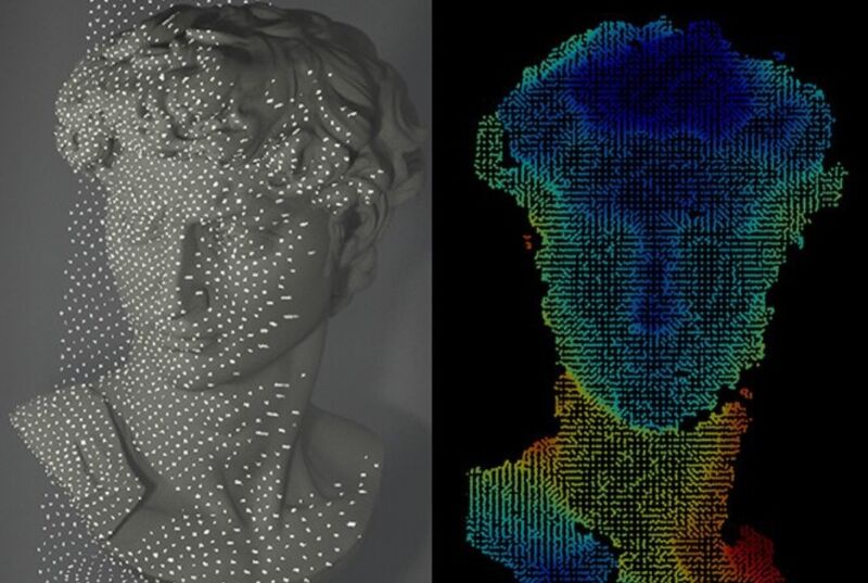
Layerless, all-at-once resin printing is a reality with the astonishing CAL system

The post Computed Axial Lithography: 3D Printing in Seconds appeared first on Make: DIY Projects and Ideas for Makers.

Anycubic Debuts Its Annual Flagship Products and Announces a New Business Department at TCT Shanghai 2024

Anycubic Debuts Its Annual Flagship Products and Announces a New Business Department at TCT Shanghai 2024

Shanghai, China – May 7th, 2024 – Anycubic, a renowned brand in consumer 3D printing, showcased its latest advancements at the highly anticipated TCT Shanghai event, held at Booth number 8E22. The event coincided with Anycubic’s Annual Product Launch Event, hosted by James Ouyang, the co-founder of Anycubic. James took the stage in front of […]

The post Anycubic Debuts Its Annual Flagship Products and Announces a New Business Department at TCT Shanghai 2024 appeared first on Make: DIY Projects and Ideas for Makers.

Anycubic Debuts Its Annual Flagship Products and Announces a New Business Department at TCT Shanghai 2024

Anycubic Debuts Its Annual Flagship Products and Announces a New Business Department at TCT Shanghai 2024

Shanghai, China – May 7th, 2024 – Anycubic, a renowned brand in consumer 3D printing, showcased its latest advancements at the highly anticipated TCT Shanghai event, held at Booth number 8E22. The event coincided with Anycubic’s Annual Product Launch Event, hosted by James Ouyang, the co-founder of Anycubic. James took the stage in front of […]

The post Anycubic Debuts Its Annual Flagship Products and Announces a New Business Department at TCT Shanghai 2024 appeared first on Make: DIY Projects and Ideas for Makers.

Walk This Way: DIY Mobility Walker

Walk This Way: DIY Mobility Walker

Mobility walkers can be expensive. Build your own personalized, strength-tested version for less.

The post Walk This Way: DIY Mobility Walker appeared first on Make: DIY Projects and Ideas for Makers.

  • ✇MIT News - Nanoscience and nanotechnology | MIT.nano
  • MIT’s tiny technologies go to WashingtonKoch Institute
    On Nov. 7, a team from the Marble Center for Cancer Nanomedicine at MIT showed a Washington audience several examples of how nanotechnologies developed at the Institute can transform the detection and treatment of cancer and other diseases. The team was one of 40 innovative groups featured at “American Possibilities: A White House Demo Day.” Technology on view spanned energy, artificial intelligence, climate, and health, highlighting advancements that contribute to building a better future for
     

MIT’s tiny technologies go to Washington

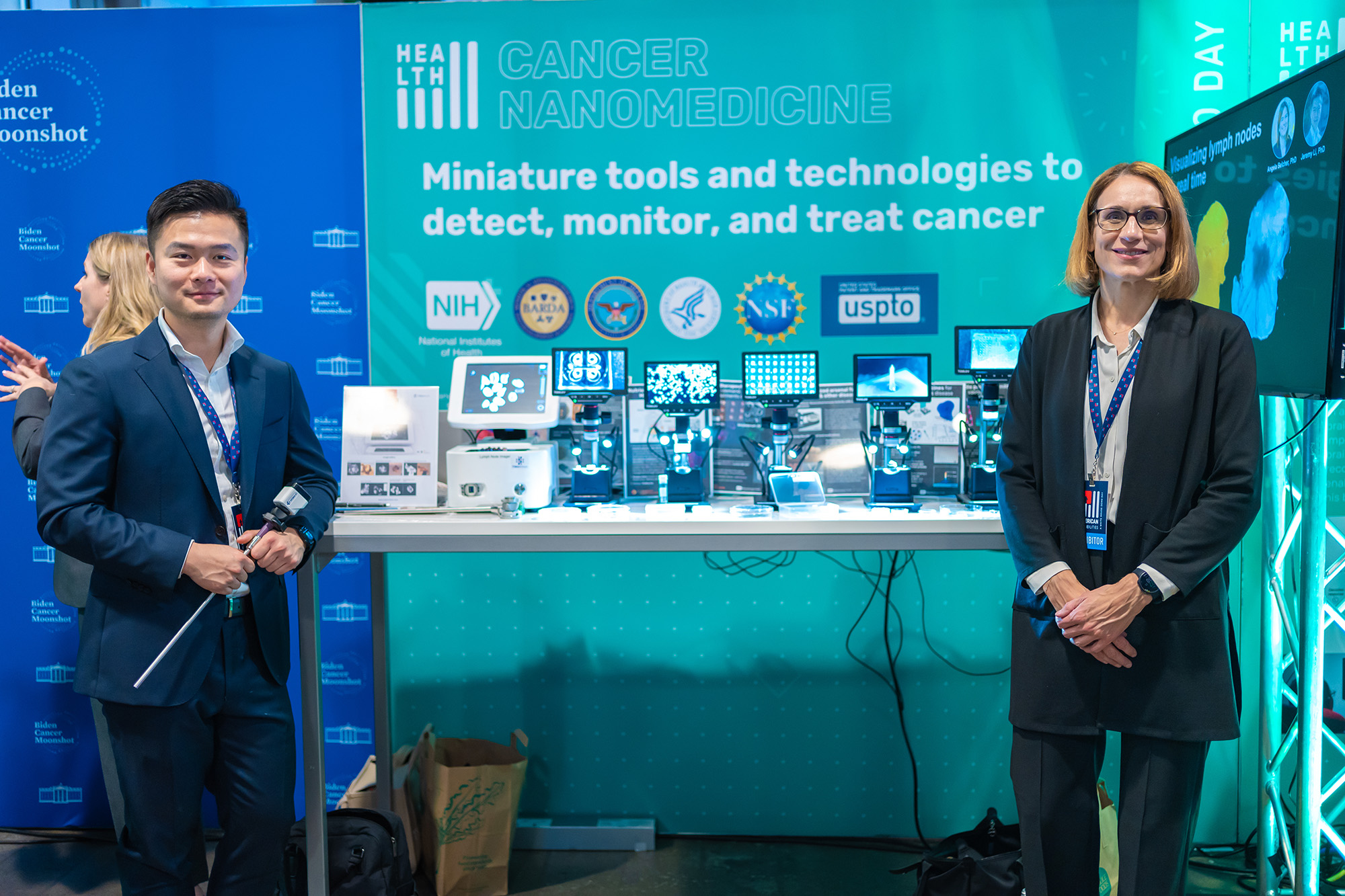
On Nov. 7, a team from the Marble Center for Cancer Nanomedicine at MIT showed a Washington audience several examples of how nanotechnologies developed at the Institute can transform the detection and treatment of cancer and other diseases.

The team was one of 40 innovative groups featured at “American Possibilities: A White House Demo Day.” Technology on view spanned energy, artificial intelligence, climate, and health, highlighting advancements that contribute to building a better future for all Americans.

Participants included President Joe Biden, Biden-Harris administration leaders and White House staff, members of Congress, federal R&D funding agencies, scientists and engineers, academics, students, and science and technology industry innovators. The event holds special significance for MIT as eight years ago, MIT's Computer Science and Artificial Intelligence Laboratory participated in the last iteration of the White House Demo Day under President Barack Obama.

“It was truly inspirational hearing from experts from all across the government, the private sector, and academia touching on so many fields,” said President Biden of the event. “It was a reminder, at least for me, of what I’ve long believed — that America can be defined by a single word... possibilities.”

Launched in 2016, the Marble Center for Cancer Nanomedicine was established at the Koch Institute for Integrative Research at MIT to serve as a hub for miniaturized biomedical technologies, especially those that address grand challenges in cancer detection, treatment, and monitoring. The center convenes Koch Institute faculty members Sangeeta Bhatia, Paula Hammond, Robert Langer, Angela Belcher, Darrell Irvine, and Daniel Anderson to advance nanomedicine, as well as to facilitate collaboration with industry partners, including Alloy Therapeutics, Danaher Corp., Fujifilm, and Sanofi. 

Ana Jaklenec, a principal research scientist at the Koch Institute, highlighted several groundbreaking technologies in vaccines and disease diagnostics and treatment at the event. Jaklenec gave demonstrations from projects from her research group, including novel vaccine formulations capable of releasing a dozen booster doses pulsed over predetermined time points, microneedle vaccine technologies, and nutrient delivery technologies for precise control over microbiome modulation and nutrient absorption.

Jaklenec describes the event as “a wonderful opportunity to meet our government leaders and policymakers and see their passion for curing cancer. But it was especially moving to interact with people representing diverse communities across the United States and hear their excitement for how our technologies could positively impact their communities.”

Jeremy Li, a former MIT postdoc, presented a technology developed in the Belcher laboratory and commercialized by the spinout Cision Vision. The startup is developing a new approach to visualize lymph nodes in real time without any injection or radiation. The shoebox-sized device was also selected as part of Time Magazine’s Best Inventions of 2023 and is currently being used in a dozen hospitals across the United States.

“It was a proud moment for Cision Vision to be part of this event and discuss our recent progress in the field of medical imaging and cancer care,” says Li, who is a co-founder and the CEO of CisionVision. “It was a humbling experience for us to hear directly from patient advocates and cancer survivors at the event. We feel more inspired than ever to bring better solutions for cancer care to patients around the world.”

Other technologies shown at the event included new approaches such as a tortoise-shaped pill designed to enhance the efficacy of oral medicines, a miniature organ-on-a-chip liver device to predict drug toxicity and model liver disease, and a wireless bioelectronic device that provides oxygen for cell therapy applications and for the treatment of chronic disease.

“The feedback from the organizers and the audience at the event has been overwhelmingly positive,” says Tarek Fadel, who led the team’s participation at the event. “Navigating the demonstration space felt like stepping into the future. As a center, we stand poised to engineer transformative tools that will truly make a difference for the future of cancer care.”

Sangeeta Bhatia, the Director of the Marble Center and the John J. and Dorothy Wilson Professor of Health Sciences and Technology and Electrical Engineering and Computer Science, adds: “The showcase of our technologies at the White House Demo Day underscores the transformative impact we aim to achieve in cancer detection and treatment. The event highlights our vision to advance cutting-edge solutions for the benefit of patients and communities worldwide.”

Ana Jaklenec (right), principal research scientist at the Koch Institute for Integrative Cancer Research at MIT, and Jeremy Li, CEO and co-founder of Cision Vision, presented at “American Possibilities: A White House Demo Day.”

Angela Belcher delivers 2023 Dresselhaus Lecture on evolving organisms for new nanomaterials

“How do we get to making nanomaterials that haven’t been evolved before?” asked Angela Belcher at the 2023 Mildred S. Dresselhaus Lecture at MIT on Nov. 20. “We can use elements that biology has already given us.”

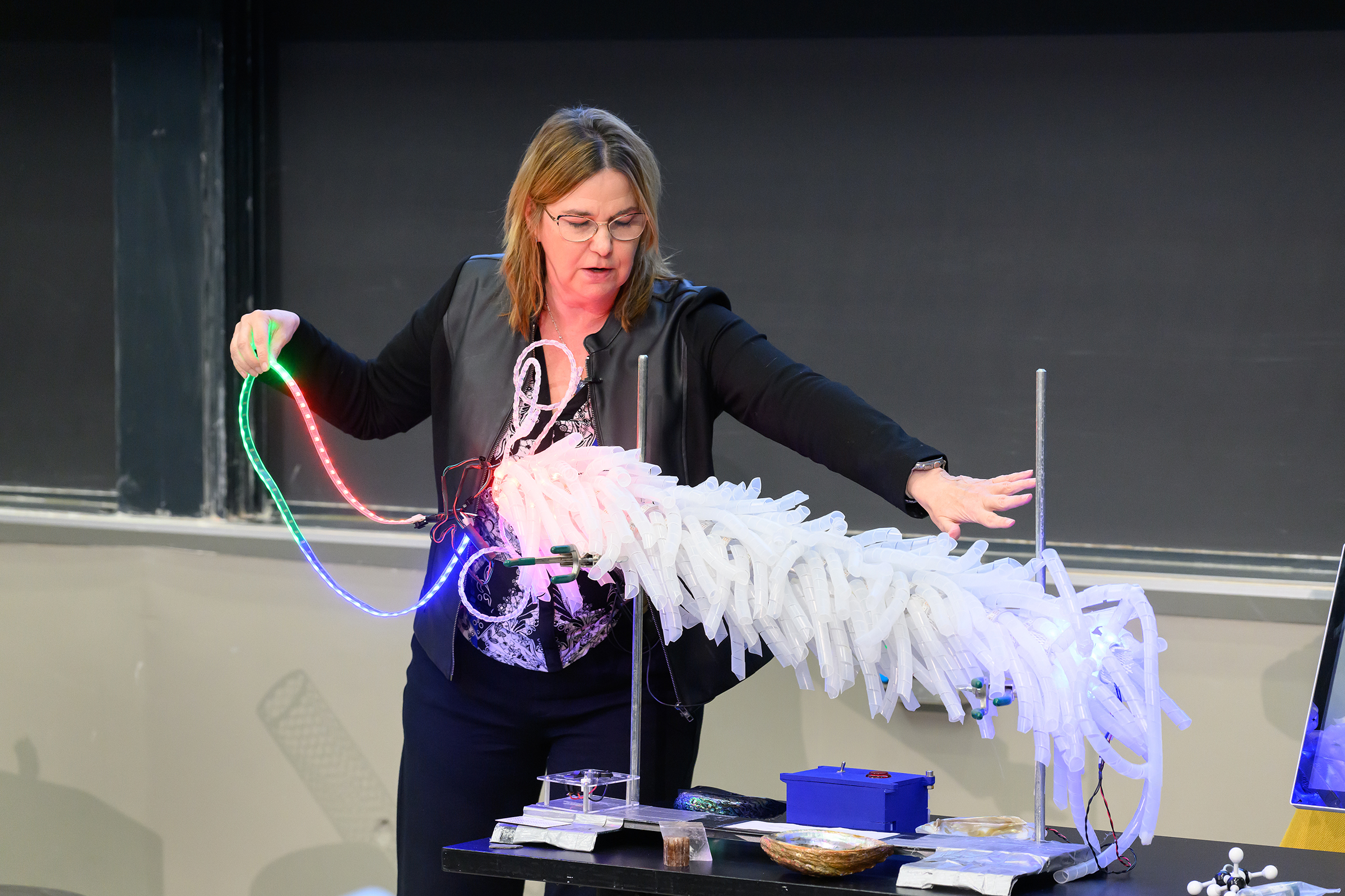
The combined in-person and virtual audience of over 300 was treated to a light-up, 3D model of M13 bacteriophage, a virus that only infects bacteria, complete with a pull-out strand of DNA. Belcher used the feather-boa-like model to show how her research group modifies the M13’s genes to add new DNA and peptide sequences to template inorganic materials.

“I love controlling materials at the nanoscale using biology,” said Belcher, the James Mason Crafts Professor of Biological Engineering, materials science professor, and of the Koch Institute of Integrative Cancer Research at MIT. “We all know if you control materials at the nanoscale and you can start to tune them, then you can have all kinds of different applications.” And the opportunities are indeed vast — from building batteries, fuel cells, and solar cells to carbon sequestration and storage, environmental remediation, catalysis, and medical diagnostics and imaging.

Belcher sprinkled her talk with models and props, lined up on a table at the front of the 10-250 lecture hall, to demonstrate a wide variety of concepts and projects made possible by the intersection of biology and nanotechnology.

Energy storage and environment

“How do you go from a DNA sequence to a functioning battery?” posed Belcher. Grabbing a model of a large carbon nanotube, she explained how her group engineered a phage to pick up carbon nanotubes that would wind all the way around the virus and then fill in with different cathode or anode materials to make nanowires for battery electrodes.

How about using the M13 bacteriophage to improve the environment? Belcher referred to a project by former student Geran Zhang PhD ’19 that proved the virus can be modified for this context, too. He used the phage to template high-surface-area, carbon-based materials that can grab small molecules and break them down, Belcher said, opening a realm of possibilities from cleaning up rivers to developing chemical warfare agents to combating smog.

Belcher’s lab worked with the U.S. Army to produce protective clothing and masks made of these carbon-based virus nanofibers. “We went from five liters in our lab to a thousand liters, then 10,000 liters in the army labs where we’re able to make kilograms of the material,” Belcher said, stressing the importance of being able to test and prototype at scale.

Imaging tools and therapeutics in cancer

In the area of biomedical imaging, Belcher explained, a lot less is known in near-infrared imaging — imaging in wavelengths above 1,000 nanometers — than other imaging techniques, yet with near-infrared scientists can see much deeper inside the body. Belcher’s lab built their own systems to image at these wavelengths. The third generation of this system provides real-time, sub-millimeter optical imaging for guided surgery.

Working with Sangeeta Bhatia, the John J. and Dorothy Wilson Professor of Engineering, Belcher used carbon nanotubes to build imaging tools that find tiny tumors during surgery that doctors otherwise would not be able to see. The tool is actually a virus engineered to carry with it a fluorescent, single-walled carbon nanotube as it seeks out the tumors.

Nearing the end of her talk, Belcher presented a goal: to develop an accessible detection and diagnostic technology for ovarian cancer in five to 10 years.

“We think that we can do it,” Belcher said. She described her students’ work developing a way to scan an entire fallopian tube, as opposed to just one small portion, to find pre-cancer lesions, and talked about the team of MIT faculty, doctors, and researchers working collectively toward this goal.

“Part of the secret of life and the meaning of life is helping other people enjoy the passage of time,” said Belcher in her closing remarks. “I think that we can all do that by working to solve some of the biggest issues on the planet, including helping to diagnose and treat ovarian cancer early so people have more time to spend with their family.”

Honoring Mildred S. Dresselhaus

Belcher was the fifth speaker to deliver the Dresselhaus Lecture, an annual event organized by MIT.nano to honor the late MIT physics and electrical engineering Institute Professor Mildred Dresselhaus. The lecture features a speaker from anywhere in the world whose leadership and impact echo Dresselhaus’s life, accomplishments, and values.

“Millie was and is a huge hero of mine,” said Belcher. “Giving a lecture in Millie’s name is just the greatest honor.”

Belcher dedicated the talk to Dresselhaus, whom she described with an array of accolades — a trailblazer, a genius, an amazing mentor, teacher, and inventor. “Just knowing her was such a privilege,” she said.

Belcher also dedicated her talk to her own grandmother and mother, both of whom passed away from cancer, as well as late MIT professors Susan Lindquist and Angelika Amon, who both died of ovarian cancer.

“I’ve been so fortunate to work with just the most talented and dedicated graduate students, undergraduate students, postdocs, and researchers,” concluded Belcher. “It has been a pure joy to be in partnership with all of you to solve these very daunting problems.”

© Photo: Justin Knight

Professor Angela Belcher pulls a single strand of DNA out of a light-up model of M13 bacteriophage, a virus that only infects bacteria. Belcher’s lab modifies the M13’s genes to add new DNA and peptide sequences to template inorganic materials.

Walk This Way: DIY Mobility Walker

Walk This Way: DIY Mobility Walker

Mobility walkers can be expensive. Build your own personalized, strength-tested version for less.

The post Walk This Way: DIY Mobility Walker appeared first on Make: DIY Projects and Ideas for Makers.

  • ✇MIT News - Nanoscience and nanotechnology | MIT.nano
  • MIT’s tiny technologies go to WashingtonKoch Institute
    On Nov. 7, a team from the Marble Center for Cancer Nanomedicine at MIT showed a Washington audience several examples of how nanotechnologies developed at the Institute can transform the detection and treatment of cancer and other diseases. The team was one of 40 innovative groups featured at “American Possibilities: A White House Demo Day.” Technology on view spanned energy, artificial intelligence, climate, and health, highlighting advancements that contribute to building a better future for
     

MIT’s tiny technologies go to Washington

On Nov. 7, a team from the Marble Center for Cancer Nanomedicine at MIT showed a Washington audience several examples of how nanotechnologies developed at the Institute can transform the detection and treatment of cancer and other diseases.

The team was one of 40 innovative groups featured at “American Possibilities: A White House Demo Day.” Technology on view spanned energy, artificial intelligence, climate, and health, highlighting advancements that contribute to building a better future for all Americans.

Participants included President Joe Biden, Biden-Harris administration leaders and White House staff, members of Congress, federal R&D funding agencies, scientists and engineers, academics, students, and science and technology industry innovators. The event holds special significance for MIT as eight years ago, MIT's Computer Science and Artificial Intelligence Laboratory participated in the last iteration of the White House Demo Day under President Barack Obama.

“It was truly inspirational hearing from experts from all across the government, the private sector, and academia touching on so many fields,” said President Biden of the event. “It was a reminder, at least for me, of what I’ve long believed — that America can be defined by a single word... possibilities.”

Launched in 2016, the Marble Center for Cancer Nanomedicine was established at the Koch Institute for Integrative Research at MIT to serve as a hub for miniaturized biomedical technologies, especially those that address grand challenges in cancer detection, treatment, and monitoring. The center convenes Koch Institute faculty members Sangeeta Bhatia, Paula Hammond, Robert Langer, Angela Belcher, Darrell Irvine, and Daniel Anderson to advance nanomedicine, as well as to facilitate collaboration with industry partners, including Alloy Therapeutics, Danaher Corp., Fujifilm, and Sanofi. 

Ana Jaklenec, a principal research scientist at the Koch Institute, highlighted several groundbreaking technologies in vaccines and disease diagnostics and treatment at the event. Jaklenec gave demonstrations from projects from her research group, including novel vaccine formulations capable of releasing a dozen booster doses pulsed over predetermined time points, microneedle vaccine technologies, and nutrient delivery technologies for precise control over microbiome modulation and nutrient absorption.

Jaklenec describes the event as “a wonderful opportunity to meet our government leaders and policymakers and see their passion for curing cancer. But it was especially moving to interact with people representing diverse communities across the United States and hear their excitement for how our technologies could positively impact their communities.”

Jeremy Li, a former MIT postdoc, presented a technology developed in the Belcher laboratory and commercialized by the spinout Cision Vision. The startup is developing a new approach to visualize lymph nodes in real time without any injection or radiation. The shoebox-sized device was also selected as part of Time Magazine’s Best Inventions of 2023 and is currently being used in a dozen hospitals across the United States.

“It was a proud moment for Cision Vision to be part of this event and discuss our recent progress in the field of medical imaging and cancer care,” says Li, who is a co-founder and the CEO of CisionVision. “It was a humbling experience for us to hear directly from patient advocates and cancer survivors at the event. We feel more inspired than ever to bring better solutions for cancer care to patients around the world.”

Other technologies shown at the event included new approaches such as a tortoise-shaped pill designed to enhance the efficacy of oral medicines, a miniature organ-on-a-chip liver device to predict drug toxicity and model liver disease, and a wireless bioelectronic device that provides oxygen for cell therapy applications and for the treatment of chronic disease.

“The feedback from the organizers and the audience at the event has been overwhelmingly positive,” says Tarek Fadel, who led the team’s participation at the event. “Navigating the demonstration space felt like stepping into the future. As a center, we stand poised to engineer transformative tools that will truly make a difference for the future of cancer care.”

Sangeeta Bhatia, the Director of the Marble Center and the John J. and Dorothy Wilson Professor of Health Sciences and Technology and Electrical Engineering and Computer Science, adds: “The showcase of our technologies at the White House Demo Day underscores the transformative impact we aim to achieve in cancer detection and treatment. The event highlights our vision to advance cutting-edge solutions for the benefit of patients and communities worldwide.”

Ana Jaklenec (right), principal research scientist at the Koch Institute for Integrative Cancer Research at MIT, and Jeremy Li, CEO and co-founder of Cision Vision, presented at “American Possibilities: A White House Demo Day.”

Angela Belcher delivers 2023 Dresselhaus Lecture on evolving organisms for new nanomaterials

“How do we get to making nanomaterials that haven’t been evolved before?” asked Angela Belcher at the 2023 Mildred S. Dresselhaus Lecture at MIT on Nov. 20. “We can use elements that biology has already given us.”

The combined in-person and virtual audience of over 300 was treated to a light-up, 3D model of M13 bacteriophage, a virus that only infects bacteria, complete with a pull-out strand of DNA. Belcher used the feather-boa-like model to show how her research group modifies the M13’s genes to add new DNA and peptide sequences to template inorganic materials.

“I love controlling materials at the nanoscale using biology,” said Belcher, the James Mason Crafts Professor of Biological Engineering, materials science professor, and of the Koch Institute of Integrative Cancer Research at MIT. “We all know if you control materials at the nanoscale and you can start to tune them, then you can have all kinds of different applications.” And the opportunities are indeed vast — from building batteries, fuel cells, and solar cells to carbon sequestration and storage, environmental remediation, catalysis, and medical diagnostics and imaging.

Belcher sprinkled her talk with models and props, lined up on a table at the front of the 10-250 lecture hall, to demonstrate a wide variety of concepts and projects made possible by the intersection of biology and nanotechnology.

Energy storage and environment

“How do you go from a DNA sequence to a functioning battery?” posed Belcher. Grabbing a model of a large carbon nanotube, she explained how her group engineered a phage to pick up carbon nanotubes that would wind all the way around the virus and then fill in with different cathode or anode materials to make nanowires for battery electrodes.

How about using the M13 bacteriophage to improve the environment? Belcher referred to a project by former student Geran Zhang PhD ’19 that proved the virus can be modified for this context, too. He used the phage to template high-surface-area, carbon-based materials that can grab small molecules and break them down, Belcher said, opening a realm of possibilities from cleaning up rivers to developing chemical warfare agents to combating smog.

Belcher’s lab worked with the U.S. Army to produce protective clothing and masks made of these carbon-based virus nanofibers. “We went from five liters in our lab to a thousand liters, then 10,000 liters in the army labs where we’re able to make kilograms of the material,” Belcher said, stressing the importance of being able to test and prototype at scale.

Imaging tools and therapeutics in cancer

In the area of biomedical imaging, Belcher explained, a lot less is known in near-infrared imaging — imaging in wavelengths above 1,000 nanometers — than other imaging techniques, yet with near-infrared scientists can see much deeper inside the body. Belcher’s lab built their own systems to image at these wavelengths. The third generation of this system provides real-time, sub-millimeter optical imaging for guided surgery.

Working with Sangeeta Bhatia, the John J. and Dorothy Wilson Professor of Engineering, Belcher used carbon nanotubes to build imaging tools that find tiny tumors during surgery that doctors otherwise would not be able to see. The tool is actually a virus engineered to carry with it a fluorescent, single-walled carbon nanotube as it seeks out the tumors.

Nearing the end of her talk, Belcher presented a goal: to develop an accessible detection and diagnostic technology for ovarian cancer in five to 10 years.

“We think that we can do it,” Belcher said. She described her students’ work developing a way to scan an entire fallopian tube, as opposed to just one small portion, to find pre-cancer lesions, and talked about the team of MIT faculty, doctors, and researchers working collectively toward this goal.

“Part of the secret of life and the meaning of life is helping other people enjoy the passage of time,” said Belcher in her closing remarks. “I think that we can all do that by working to solve some of the biggest issues on the planet, including helping to diagnose and treat ovarian cancer early so people have more time to spend with their family.”

Honoring Mildred S. Dresselhaus

Belcher was the fifth speaker to deliver the Dresselhaus Lecture, an annual event organized by MIT.nano to honor the late MIT physics and electrical engineering Institute Professor Mildred Dresselhaus. The lecture features a speaker from anywhere in the world whose leadership and impact echo Dresselhaus’s life, accomplishments, and values.

“Millie was and is a huge hero of mine,” said Belcher. “Giving a lecture in Millie’s name is just the greatest honor.”

Belcher dedicated the talk to Dresselhaus, whom she described with an array of accolades — a trailblazer, a genius, an amazing mentor, teacher, and inventor. “Just knowing her was such a privilege,” she said.

Belcher also dedicated her talk to her own grandmother and mother, both of whom passed away from cancer, as well as late MIT professors Susan Lindquist and Angelika Amon, who both died of ovarian cancer.

“I’ve been so fortunate to work with just the most talented and dedicated graduate students, undergraduate students, postdocs, and researchers,” concluded Belcher. “It has been a pure joy to be in partnership with all of you to solve these very daunting problems.”

© Photo: Justin Knight

Professor Angela Belcher pulls a single strand of DNA out of a light-up model of M13 bacteriophage, a virus that only infects bacteria. Belcher’s lab modifies the M13’s genes to add new DNA and peptide sequences to template inorganic materials.
  • ✇Make: DIY Projects and Ideas for Makers
  • Staggering Layers For Added Strength In Your 3D PrintsCaleb Kraft
    The constant hunt for more efficient and useful ways to use these 3d printers keeps turning up interesting results. For quite some time, people have speculated on various methods of arranging our 3d print layers for better adhesion and stronger results. Stefan from CNC Kitchen tackles the subject to see if there’s any validity to […] The post Staggering Layers For Added Strength In Your 3D Prints appeared first on Make: DIY Projects and Ideas for Makers.
     

Staggering Layers For Added Strength In Your 3D Prints

Staggering Layers For Added Strength In Your 3D Prints

The constant hunt for more efficient and useful ways to use these 3d printers keeps turning up interesting results. For quite some time, people have speculated on various methods of arranging our 3d print layers for better adhesion and stronger results. Stefan from CNC Kitchen tackles the subject to see if there’s any validity to […]

The post Staggering Layers For Added Strength In Your 3D Prints appeared first on Make: DIY Projects and Ideas for Makers.

Can I Use AI To Make Models For 3D Printing?

Can I Use AI To Make Models For 3D Printing?

Kind of. Text-to-3D-model generators are here, but not optimized for printing

The post Can I Use AI To Make Models For 3D Printing? appeared first on Make: DIY Projects and Ideas for Makers.

  • ✇MIT News - Nanoscience and nanotechnology | MIT.nano
  • MIT’s tiny technologies go to WashingtonKoch Institute
    On Nov. 7, a team from the Marble Center for Cancer Nanomedicine at MIT showed a Washington audience several examples of how nanotechnologies developed at the Institute can transform the detection and treatment of cancer and other diseases. The team was one of 40 innovative groups featured at “American Possibilities: A White House Demo Day.” Technology on view spanned energy, artificial intelligence, climate, and health, highlighting advancements that contribute to building a better future for
     

MIT’s tiny technologies go to Washington

On Nov. 7, a team from the Marble Center for Cancer Nanomedicine at MIT showed a Washington audience several examples of how nanotechnologies developed at the Institute can transform the detection and treatment of cancer and other diseases.

The team was one of 40 innovative groups featured at “American Possibilities: A White House Demo Day.” Technology on view spanned energy, artificial intelligence, climate, and health, highlighting advancements that contribute to building a better future for all Americans.

Participants included President Joe Biden, Biden-Harris administration leaders and White House staff, members of Congress, federal R&D funding agencies, scientists and engineers, academics, students, and science and technology industry innovators. The event holds special significance for MIT as eight years ago, MIT's Computer Science and Artificial Intelligence Laboratory participated in the last iteration of the White House Demo Day under President Barack Obama.

“It was truly inspirational hearing from experts from all across the government, the private sector, and academia touching on so many fields,” said President Biden of the event. “It was a reminder, at least for me, of what I’ve long believed — that America can be defined by a single word... possibilities.”

Launched in 2016, the Marble Center for Cancer Nanomedicine was established at the Koch Institute for Integrative Research at MIT to serve as a hub for miniaturized biomedical technologies, especially those that address grand challenges in cancer detection, treatment, and monitoring. The center convenes Koch Institute faculty members Sangeeta Bhatia, Paula Hammond, Robert Langer, Angela Belcher, Darrell Irvine, and Daniel Anderson to advance nanomedicine, as well as to facilitate collaboration with industry partners, including Alloy Therapeutics, Danaher Corp., Fujifilm, and Sanofi. 

Ana Jaklenec, a principal research scientist at the Koch Institute, highlighted several groundbreaking technologies in vaccines and disease diagnostics and treatment at the event. Jaklenec gave demonstrations from projects from her research group, including novel vaccine formulations capable of releasing a dozen booster doses pulsed over predetermined time points, microneedle vaccine technologies, and nutrient delivery technologies for precise control over microbiome modulation and nutrient absorption.

Jaklenec describes the event as “a wonderful opportunity to meet our government leaders and policymakers and see their passion for curing cancer. But it was especially moving to interact with people representing diverse communities across the United States and hear their excitement for how our technologies could positively impact their communities.”

Jeremy Li, a former MIT postdoc, presented a technology developed in the Belcher laboratory and commercialized by the spinout Cision Vision. The startup is developing a new approach to visualize lymph nodes in real time without any injection or radiation. The shoebox-sized device was also selected as part of Time Magazine’s Best Inventions of 2023 and is currently being used in a dozen hospitals across the United States.

“It was a proud moment for Cision Vision to be part of this event and discuss our recent progress in the field of medical imaging and cancer care,” says Li, who is a co-founder and the CEO of CisionVision. “It was a humbling experience for us to hear directly from patient advocates and cancer survivors at the event. We feel more inspired than ever to bring better solutions for cancer care to patients around the world.”

Other technologies shown at the event included new approaches such as a tortoise-shaped pill designed to enhance the efficacy of oral medicines, a miniature organ-on-a-chip liver device to predict drug toxicity and model liver disease, and a wireless bioelectronic device that provides oxygen for cell therapy applications and for the treatment of chronic disease.

“The feedback from the organizers and the audience at the event has been overwhelmingly positive,” says Tarek Fadel, who led the team’s participation at the event. “Navigating the demonstration space felt like stepping into the future. As a center, we stand poised to engineer transformative tools that will truly make a difference for the future of cancer care.”

Sangeeta Bhatia, the Director of the Marble Center and the John J. and Dorothy Wilson Professor of Health Sciences and Technology and Electrical Engineering and Computer Science, adds: “The showcase of our technologies at the White House Demo Day underscores the transformative impact we aim to achieve in cancer detection and treatment. The event highlights our vision to advance cutting-edge solutions for the benefit of patients and communities worldwide.”

Ana Jaklenec (right), principal research scientist at the Koch Institute for Integrative Cancer Research at MIT, and Jeremy Li, CEO and co-founder of Cision Vision, presented at “American Possibilities: A White House Demo Day.”

Angela Belcher delivers 2023 Dresselhaus Lecture on evolving organisms for new nanomaterials

“How do we get to making nanomaterials that haven’t been evolved before?” asked Angela Belcher at the 2023 Mildred S. Dresselhaus Lecture at MIT on Nov. 20. “We can use elements that biology has already given us.”

The combined in-person and virtual audience of over 300 was treated to a light-up, 3D model of M13 bacteriophage, a virus that only infects bacteria, complete with a pull-out strand of DNA. Belcher used the feather-boa-like model to show how her research group modifies the M13’s genes to add new DNA and peptide sequences to template inorganic materials.

“I love controlling materials at the nanoscale using biology,” said Belcher, the James Mason Crafts Professor of Biological Engineering, materials science professor, and of the Koch Institute of Integrative Cancer Research at MIT. “We all know if you control materials at the nanoscale and you can start to tune them, then you can have all kinds of different applications.” And the opportunities are indeed vast — from building batteries, fuel cells, and solar cells to carbon sequestration and storage, environmental remediation, catalysis, and medical diagnostics and imaging.

Belcher sprinkled her talk with models and props, lined up on a table at the front of the 10-250 lecture hall, to demonstrate a wide variety of concepts and projects made possible by the intersection of biology and nanotechnology.

Energy storage and environment

“How do you go from a DNA sequence to a functioning battery?” posed Belcher. Grabbing a model of a large carbon nanotube, she explained how her group engineered a phage to pick up carbon nanotubes that would wind all the way around the virus and then fill in with different cathode or anode materials to make nanowires for battery electrodes.

How about using the M13 bacteriophage to improve the environment? Belcher referred to a project by former student Geran Zhang PhD ’19 that proved the virus can be modified for this context, too. He used the phage to template high-surface-area, carbon-based materials that can grab small molecules and break them down, Belcher said, opening a realm of possibilities from cleaning up rivers to developing chemical warfare agents to combating smog.

Belcher’s lab worked with the U.S. Army to produce protective clothing and masks made of these carbon-based virus nanofibers. “We went from five liters in our lab to a thousand liters, then 10,000 liters in the army labs where we’re able to make kilograms of the material,” Belcher said, stressing the importance of being able to test and prototype at scale.

Imaging tools and therapeutics in cancer

In the area of biomedical imaging, Belcher explained, a lot less is known in near-infrared imaging — imaging in wavelengths above 1,000 nanometers — than other imaging techniques, yet with near-infrared scientists can see much deeper inside the body. Belcher’s lab built their own systems to image at these wavelengths. The third generation of this system provides real-time, sub-millimeter optical imaging for guided surgery.

Working with Sangeeta Bhatia, the John J. and Dorothy Wilson Professor of Engineering, Belcher used carbon nanotubes to build imaging tools that find tiny tumors during surgery that doctors otherwise would not be able to see. The tool is actually a virus engineered to carry with it a fluorescent, single-walled carbon nanotube as it seeks out the tumors.

Nearing the end of her talk, Belcher presented a goal: to develop an accessible detection and diagnostic technology for ovarian cancer in five to 10 years.

“We think that we can do it,” Belcher said. She described her students’ work developing a way to scan an entire fallopian tube, as opposed to just one small portion, to find pre-cancer lesions, and talked about the team of MIT faculty, doctors, and researchers working collectively toward this goal.

“Part of the secret of life and the meaning of life is helping other people enjoy the passage of time,” said Belcher in her closing remarks. “I think that we can all do that by working to solve some of the biggest issues on the planet, including helping to diagnose and treat ovarian cancer early so people have more time to spend with their family.”

Honoring Mildred S. Dresselhaus

Belcher was the fifth speaker to deliver the Dresselhaus Lecture, an annual event organized by MIT.nano to honor the late MIT physics and electrical engineering Institute Professor Mildred Dresselhaus. The lecture features a speaker from anywhere in the world whose leadership and impact echo Dresselhaus’s life, accomplishments, and values.

“Millie was and is a huge hero of mine,” said Belcher. “Giving a lecture in Millie’s name is just the greatest honor.”

Belcher dedicated the talk to Dresselhaus, whom she described with an array of accolades — a trailblazer, a genius, an amazing mentor, teacher, and inventor. “Just knowing her was such a privilege,” she said.

Belcher also dedicated her talk to her own grandmother and mother, both of whom passed away from cancer, as well as late MIT professors Susan Lindquist and Angelika Amon, who both died of ovarian cancer.

“I’ve been so fortunate to work with just the most talented and dedicated graduate students, undergraduate students, postdocs, and researchers,” concluded Belcher. “It has been a pure joy to be in partnership with all of you to solve these very daunting problems.”

© Photo: Justin Knight

Professor Angela Belcher pulls a single strand of DNA out of a light-up model of M13 bacteriophage, a virus that only infects bacteria. Belcher’s lab modifies the M13’s genes to add new DNA and peptide sequences to template inorganic materials.
  • ✇MIT News - Nanoscience and nanotechnology | MIT.nano
  • Using AI to optimize for rapid neural imagingRachel Gordon | MIT CSAIL
    Connectomics, the ambitious field of study that seeks to map the intricate network of animal brains, is undergoing a growth spurt. Within the span of a decade, it has journeyed from its nascent stages to a discipline that is poised to (hopefully) unlock the enigmas of cognition and the physical underpinning of neuropathologies such as in Alzheimer’s disease.  At its forefront is the use of powerful electron microscopes, which researchers from the MIT Computer Science and Artificial Intelligence
     

Using AI to optimize for rapid neural imaging

Connectomics, the ambitious field of study that seeks to map the intricate network of animal brains, is undergoing a growth spurt. Within the span of a decade, it has journeyed from its nascent stages to a discipline that is poised to (hopefully) unlock the enigmas of cognition and the physical underpinning of neuropathologies such as in Alzheimer’s disease. 

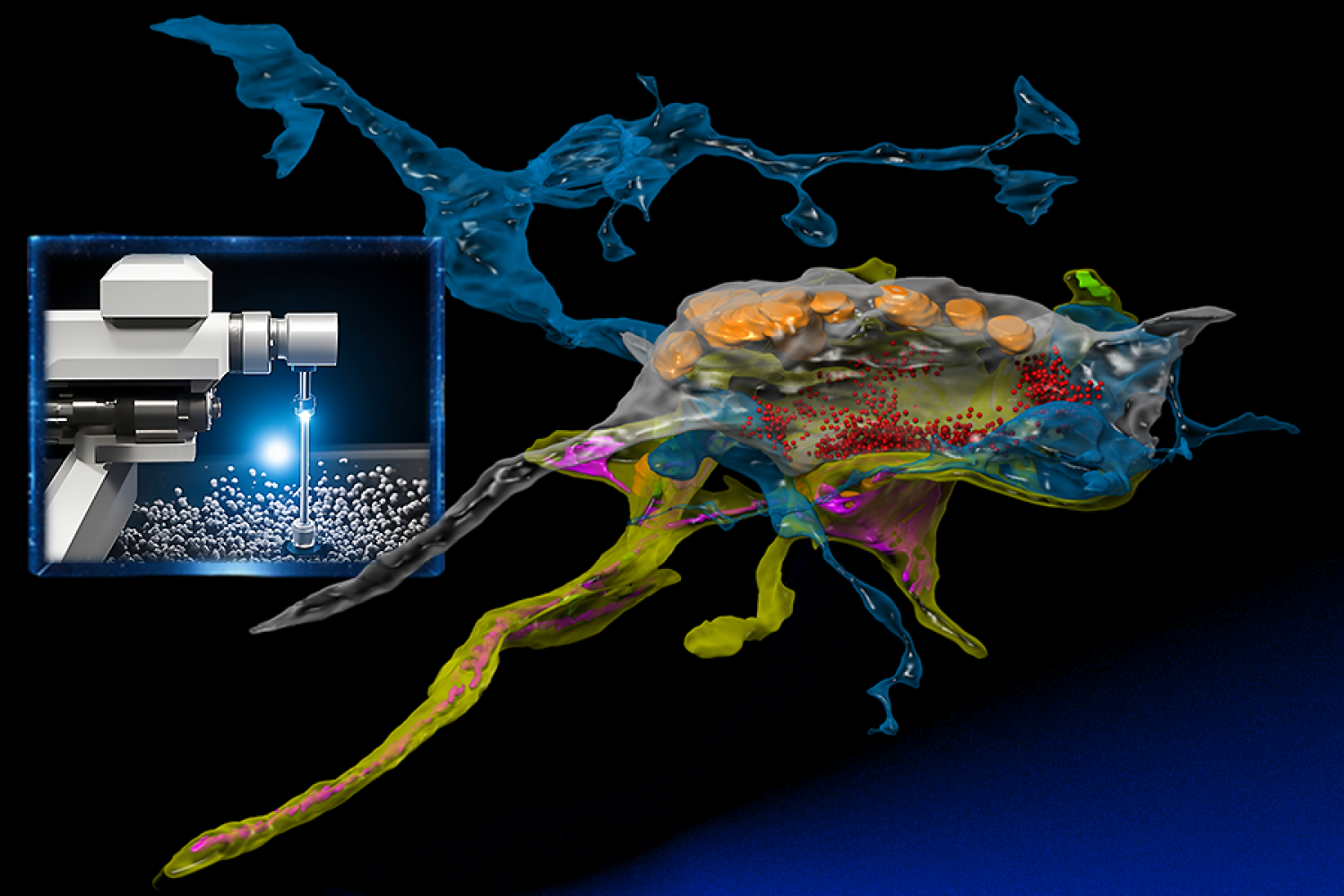
At its forefront is the use of powerful electron microscopes, which researchers from the MIT Computer Science and Artificial Intelligence Laboratory (CSAIL) and the Samuel and Lichtman Labs of Harvard University bestowed with the analytical prowess of machine learning. Unlike traditional electron microscopy, the integrated AI serves as a “brain” that learns a specimen while acquiring the images, and intelligently focuses on the relevant pixels at nanoscale resolution similar to how animals inspect their worlds. 

SmartEM” assists connectomics in quickly examining and reconstructing the brain’s complex network of synapses and neurons with nanometer precision. Unlike traditional electron microscopy, its integrated AI opens new doors to understand the brain's intricate architecture.

The integration of hardware and software in the process is crucial. The team embedded a GPU into the support computer connected to their microscope. This enabled running machine-learning models on the images, helping the microscope beam be directed to areas deemed interesting by the AI. “This lets the microscope dwell longer in areas that are harder to understand until it captures what it needs,” says MIT professor and CSAIL principal investigator Nir Shavit. “This step helps in mirroring human eye control, enabling rapid understanding of the images.” 

“When we look at a human face, our eyes swiftly navigate to the focal points that deliver vital cues for effective communication and comprehension,” says the lead architect of SmartEM, Yaron Meirovitch, a visiting scientist at MIT CSAIL who is also a former postdoc and current research associate neuroscientist at Harvard. “When we immerse ourselves in a book, we don't scan all of the empty space; rather, we direct our gaze towards the words and characters with ambiguity relative to our sentence expectations. This phenomenon within the human visual system has paved the way for the birth of the novel microscope concept.” 

For the task of reconstructing a human brain segment of about 100,000 neurons, achieving this with a conventional microscope would necessitate a decade of continuous imaging and a prohibitive budget. However, with SmartEM, by investing in four of these innovative microscopes at less than $1 million each, the task could be completed in a mere three months.

Nobel Prizes and little worms  

Over a century ago, Spanish neuroscientist Santiago Ramón y Cajal was heralded as being the first to characterize the structure of the nervous system. Employing the rudimentary light microscopes of his time, he embarked on leading explorations into neuroscience, laying the foundational understanding of neurons and sketching the initial outlines of this expansive and uncharted realm — a feat that earned him a Nobel Prize. He noted, on the topics of inspiration and discovery, that “As long as our brain is a mystery, the universe, the reflection of the structure of the brain will also be a mystery.”

Progressing from these early stages, the field has advanced dramatically, evidenced by efforts in the 1980s, mapping the relatively simpler connectome of C. elegans, small worms, to today’s endeavors probing into more intricate brains of organisms like zebrafish and mice. This evolution reflects not only enormous strides, but also escalating complexities and demands: mapping the mouse brain alone means managing a staggering thousand petabytes of data, a task that vastly eclipses the storage capabilities of any university, the team says. 

Testing the waters

For their own work, Meirovitch and others from the research team studied 30-nanometer thick slices of octopus tissue that were mounted on tapes, put on wafers, and finally inserted into the electron microscopes. Each section of an octopus brain, comprising billions of pixels, was imaged, letting the scientists reconstruct the slices into a three-dimensional cube at nanometer resolution. This provided an ultra-detailed view of synapses. The chief aim? To colorize these images, identify each neuron, and understand their interrelationships, thereby creating a detailed map or “connectome” of the brain's circuitry.

“SmartEM will cut the imaging time of such projects from two weeks to 1.5 days,” says Meirovitch. “Neuroscience labs that currently can't be engaged with expensive and long EM imaging will be able to do it now,” The method should also allow synapse-level circuit analysis in samples from patients with psychiatric and neurologic disorders. 

Down the line, the team envisions a future where connectomics is both affordable and accessible. They hope that with tools like SmartEM, a wider spectrum of research institutions could contribute to neuroscience without relying on large partnerships, and that the method will soon be a standard pipeline in cases where biopsies from living patients are available. Additionally, they’re eager to apply the tech to understand pathologies, extending utility beyond just connectomics. “We are now endeavoring to introduce this to hospitals for large biopsies, utilizing electron microscopes, aiming to make pathology studies more efficient,” says Shavit. 

Two other authors on the paper have MIT CSAIL ties: lead author Lu Mi MCS ’19, PhD ’22, who is now a postdoc at the Allen Institute for Brain Science, and Shashata Sawmya, an MIT graduate student in the lab. The other lead authors are Core Francisco Park and Pavel Potocek, while Harvard professors Jeff Lichtman and Aravi Samuel are additional senior authors. Their research was supported by the NIH BRAIN Initiative and was presented at the 2023 International Conference on Machine Learning (ICML) Workshop on Computational Biology. The work was done in collaboration with scientists from Thermo Fisher Scientific.

© Left image: Yaron Meirovitch via the Stable Diffusion XL AI image generator and Alex Shipps via the Midjourney AI image generator. Right image: Daniel Berger and Meirovitch, edited by Alex Shipps/MIT CSAIL

MIT researchers invented a technology and software to take electron microscopy to the next level by seamlessly integrating real-time machine learning into the imaging process — “smart microscopy.”

Pixel-by-pixel analysis yields insights into lithium-ion batteries

By mining data from X-ray images, researchers at MIT, Stanford University, SLAC National Accelerator, and the Toyota Research Institute have made significant new discoveries about the reactivity of lithium iron phosphate, a material used in batteries for electric cars and in other rechargeable batteries.

The new technique has revealed several phenomena that were previously impossible to see, including variations in the rate of lithium intercalation reactions in different regions of a lithium iron phosphate nanoparticle.

The paper’s most significant practical finding — that these variations in reaction rate are correlated with differences in the thickness of the carbon coating on the surface of the particles — could lead to improvements in the efficiency of charging and discharging such batteries.

“What we learned from this study is that it’s the interfaces that really control the dynamics of the battery, especially in today’s modern batteries made from nanoparticles of the active material. That means that our focus should really be on engineering that interface,” says Martin Bazant, the E.G. Roos Professor of Chemical Engineering and a professor of mathematics at MIT, who is the senior author of the study.

63 pairs of almond-shaped iron phosphate particles colored red, green, and yellow are pictured upon black background. Each pair has a pointer below with green center that stays still as the red end spins clockwise.

This approach to discovering the physics behind complex patterns in images could also be used to gain insights into many other materials, not only other types of batteries but also biological systems, such as dividing cells in a developing embryo.

“What I find most exciting about this work is the ability to take images of a system that’s undergoing the formation of some pattern, and learning the principles that govern that,” Bazant says.

Hongbo Zhao PhD ’21, a former MIT graduate student who is now a postdoc at Princeton University, is the lead author of the new study, which appears today in Nature. Other authors include Richard Bratz, the Edwin R. Gilliland Professor of Chemical Engineering at MIT; William Chueh, an associate professor of materials science and engineering at Stanford and director of the SLAC-Stanford Battery Center; and Brian Storey, senior director of Energy and Materials at the Toyota Research Institute.

“Until now, we could make these beautiful X-ray movies of battery nanoparticles at work, but it was challenging to measure and understand subtle details of how they function because the movies were so information-rich,” Chueh says. “By applying image learning to these nanoscale movies, we can extract insights that were not previously possible.”

Modeling reaction rates

Lithium iron phosphate battery electrodes are made of many tiny particles of lithium iron phosphate, surrounded by an electrolyte solution. A typical particle is about 1 micron in diameter and about 100 nanometers thick. When the battery discharges, lithium ions flow from the electrolyte solution into the material by an electrochemical reaction known as ion intercalation. When the battery charges, the intercalation reaction is reversed, and ions flow in the opposite direction.

“Lithium iron phosphate (LFP) is an important battery material due to low cost, a good safety record, and its use of abundant elements,” Storey says. “We are seeing an increased use of LFP in the EV market, so the timing of this study could not be better.”

Before the current study, Bazant had done a great deal of theoretical modeling of patterns formed by lithium-ion intercalation. Lithium iron phosphate prefers to exist in one of two stable phases: either full of lithium ions or empty. Since 2005, Bazant has been working on mathematical models of this phenomenon, known as phase separation, which generates distinctive patterns of lithium-ion flow driven by intercalation reactions. In 2015, while on sabbatical at Stanford, he began working with Chueh to try to interpret images of lithium iron phosphate particles from scanning transmission X-ray microscopy.

Using this type of microscopy, the researchers can obtain images that reveal the concentration of lithium ions, pixel-by-pixel, at every point in the particle. They can scan the particles several times as the particles charge or discharge, allowing them to create movies of how lithium ions flow in and out of the particles.

In 2017, Bazant and his colleagues at SLAC received funding from the Toyota Research Institute to pursue further studies using this approach, along with other battery-related research projects.

By analyzing X-ray images of 63 lithium iron phosphate particles as they charged and discharged, the researchers found that the movement of lithium ions within the material could be nearly identical to the computer simulations that Bazant had created earlier. Using all 180,000 pixels as measurements, the researchers trained the computational model to produce equations that accurately describe the nonequilibrium thermodynamics and reaction kinetics of the battery material.

“Every little pixel in there is jumping from full to empty, full to empty. And we’re mapping that whole process, using our equations to understand how that’s happening,” Bazant says.

The researchers also found that the patterns of lithium-ion flow that they observed could reveal spatial variations in the rate at which lithium ions are absorbed at each location on the particle surface.

“It was a real surprise to us that we could learn the heterogeneities in the system — in this case, the variations in surface reaction rate — simply by looking at the images,” Bazant says. “There are regions that seem to be fast and others that seem to be slow.”

Furthermore, the researchers showed that these differences in reaction rate were correlated with the thickness of the carbon coating on the surface of the lithium iron phosphate particles. That carbon coating is applied to lithium iron phosphate to help it conduct electricity — otherwise the material would conduct too slowly to be useful as a battery.

“We discovered at the nano scale that variation of the carbon coating thickness directly controls the rate, which is something you could never figure out if you didn't have all of this modeling and image analysis,” Bazant says.

The findings also offer quantitative support for a hypothesis Bazant formulated several years ago: that the performance of lithium iron phosphate electrodes is limited primarily by the rate of coupled ion-electron transfer at the interface between the solid particle and the carbon coating, rather than the rate of lithium-ion diffusion in the solid.

Optimized materials

The results from this study suggest that optimizing the thickness of the carbon layer on the electrode surface could help researchers to design batteries that would work more efficiently, the researchers say.

“This is the first study that's been able to directly attribute a property of the battery material with a physical property of the coating,” Bazant says. “The focus for optimizing and designing batteries should be on controlling reaction kinetics at the interface of the electrolyte and electrode.”

“This publication is the culmination of six years of dedication and collaboration,” Storey says. “This technique allows us to unlock the inner workings of the battery in a way not previously possible. Our next goal is to improve battery design by applying this new understanding.”  

In addition to using this type of analysis on other battery materials, Bazant anticipates that it could be useful for studying pattern formation in other chemical and biological systems.

This work was supported by the Toyota Research Institute through the Accelerated Materials Design and Discovery program.

© Courtesy of the researchers

By mining X-ray images, MIT researchers have made significant new discoveries about the reactivity of lithium iron phosphate, a material used in batteries for electric cars and in other rechargeable batteries. In each pair pictured, actual particles are on the left and the researchers’ simulations are on the right.

Can I Use AI To Make Models For 3D Printing?

Can I Use AI To Make Models For 3D Printing?

Kind of. Text-to-3D-model generators are here, but not optimized for printing

The post Can I Use AI To Make Models For 3D Printing? appeared first on Make: DIY Projects and Ideas for Makers.

  • ✇MIT News - Nanoscience and nanotechnology | MIT.nano
  • MIT’s tiny technologies go to WashingtonKoch Institute
    On Nov. 7, a team from the Marble Center for Cancer Nanomedicine at MIT showed a Washington audience several examples of how nanotechnologies developed at the Institute can transform the detection and treatment of cancer and other diseases. The team was one of 40 innovative groups featured at “American Possibilities: A White House Demo Day.” Technology on view spanned energy, artificial intelligence, climate, and health, highlighting advancements that contribute to building a better future for
     

MIT’s tiny technologies go to Washington

On Nov. 7, a team from the Marble Center for Cancer Nanomedicine at MIT showed a Washington audience several examples of how nanotechnologies developed at the Institute can transform the detection and treatment of cancer and other diseases.

The team was one of 40 innovative groups featured at “American Possibilities: A White House Demo Day.” Technology on view spanned energy, artificial intelligence, climate, and health, highlighting advancements that contribute to building a better future for all Americans.

Participants included President Joe Biden, Biden-Harris administration leaders and White House staff, members of Congress, federal R&D funding agencies, scientists and engineers, academics, students, and science and technology industry innovators. The event holds special significance for MIT as eight years ago, MIT's Computer Science and Artificial Intelligence Laboratory participated in the last iteration of the White House Demo Day under President Barack Obama.

“It was truly inspirational hearing from experts from all across the government, the private sector, and academia touching on so many fields,” said President Biden of the event. “It was a reminder, at least for me, of what I’ve long believed — that America can be defined by a single word... possibilities.”

Launched in 2016, the Marble Center for Cancer Nanomedicine was established at the Koch Institute for Integrative Research at MIT to serve as a hub for miniaturized biomedical technologies, especially those that address grand challenges in cancer detection, treatment, and monitoring. The center convenes Koch Institute faculty members Sangeeta Bhatia, Paula Hammond, Robert Langer, Angela Belcher, Darrell Irvine, and Daniel Anderson to advance nanomedicine, as well as to facilitate collaboration with industry partners, including Alloy Therapeutics, Danaher Corp., Fujifilm, and Sanofi. 

Ana Jaklenec, a principal research scientist at the Koch Institute, highlighted several groundbreaking technologies in vaccines and disease diagnostics and treatment at the event. Jaklenec gave demonstrations from projects from her research group, including novel vaccine formulations capable of releasing a dozen booster doses pulsed over predetermined time points, microneedle vaccine technologies, and nutrient delivery technologies for precise control over microbiome modulation and nutrient absorption.

Jaklenec describes the event as “a wonderful opportunity to meet our government leaders and policymakers and see their passion for curing cancer. But it was especially moving to interact with people representing diverse communities across the United States and hear their excitement for how our technologies could positively impact their communities.”

Jeremy Li, a former MIT postdoc, presented a technology developed in the Belcher laboratory and commercialized by the spinout Cision Vision. The startup is developing a new approach to visualize lymph nodes in real time without any injection or radiation. The shoebox-sized device was also selected as part of Time Magazine’s Best Inventions of 2023 and is currently being used in a dozen hospitals across the United States.

“It was a proud moment for Cision Vision to be part of this event and discuss our recent progress in the field of medical imaging and cancer care,” says Li, who is a co-founder and the CEO of CisionVision. “It was a humbling experience for us to hear directly from patient advocates and cancer survivors at the event. We feel more inspired than ever to bring better solutions for cancer care to patients around the world.”

Other technologies shown at the event included new approaches such as a tortoise-shaped pill designed to enhance the efficacy of oral medicines, a miniature organ-on-a-chip liver device to predict drug toxicity and model liver disease, and a wireless bioelectronic device that provides oxygen for cell therapy applications and for the treatment of chronic disease.

“The feedback from the organizers and the audience at the event has been overwhelmingly positive,” says Tarek Fadel, who led the team’s participation at the event. “Navigating the demonstration space felt like stepping into the future. As a center, we stand poised to engineer transformative tools that will truly make a difference for the future of cancer care.”

Sangeeta Bhatia, the Director of the Marble Center and the John J. and Dorothy Wilson Professor of Health Sciences and Technology and Electrical Engineering and Computer Science, adds: “The showcase of our technologies at the White House Demo Day underscores the transformative impact we aim to achieve in cancer detection and treatment. The event highlights our vision to advance cutting-edge solutions for the benefit of patients and communities worldwide.”

Ana Jaklenec (right), principal research scientist at the Koch Institute for Integrative Cancer Research at MIT, and Jeremy Li, CEO and co-founder of Cision Vision, presented at “American Possibilities: A White House Demo Day.”

Angela Belcher delivers 2023 Dresselhaus Lecture on evolving organisms for new nanomaterials

“How do we get to making nanomaterials that haven’t been evolved before?” asked Angela Belcher at the 2023 Mildred S. Dresselhaus Lecture at MIT on Nov. 20. “We can use elements that biology has already given us.”

The combined in-person and virtual audience of over 300 was treated to a light-up, 3D model of M13 bacteriophage, a virus that only infects bacteria, complete with a pull-out strand of DNA. Belcher used the feather-boa-like model to show how her research group modifies the M13’s genes to add new DNA and peptide sequences to template inorganic materials.

“I love controlling materials at the nanoscale using biology,” said Belcher, the James Mason Crafts Professor of Biological Engineering, materials science professor, and of the Koch Institute of Integrative Cancer Research at MIT. “We all know if you control materials at the nanoscale and you can start to tune them, then you can have all kinds of different applications.” And the opportunities are indeed vast — from building batteries, fuel cells, and solar cells to carbon sequestration and storage, environmental remediation, catalysis, and medical diagnostics and imaging.

Belcher sprinkled her talk with models and props, lined up on a table at the front of the 10-250 lecture hall, to demonstrate a wide variety of concepts and projects made possible by the intersection of biology and nanotechnology.

Energy storage and environment

“How do you go from a DNA sequence to a functioning battery?” posed Belcher. Grabbing a model of a large carbon nanotube, she explained how her group engineered a phage to pick up carbon nanotubes that would wind all the way around the virus and then fill in with different cathode or anode materials to make nanowires for battery electrodes.

How about using the M13 bacteriophage to improve the environment? Belcher referred to a project by former student Geran Zhang PhD ’19 that proved the virus can be modified for this context, too. He used the phage to template high-surface-area, carbon-based materials that can grab small molecules and break them down, Belcher said, opening a realm of possibilities from cleaning up rivers to developing chemical warfare agents to combating smog.

Belcher’s lab worked with the U.S. Army to produce protective clothing and masks made of these carbon-based virus nanofibers. “We went from five liters in our lab to a thousand liters, then 10,000 liters in the army labs where we’re able to make kilograms of the material,” Belcher said, stressing the importance of being able to test and prototype at scale.

Imaging tools and therapeutics in cancer

In the area of biomedical imaging, Belcher explained, a lot less is known in near-infrared imaging — imaging in wavelengths above 1,000 nanometers — than other imaging techniques, yet with near-infrared scientists can see much deeper inside the body. Belcher’s lab built their own systems to image at these wavelengths. The third generation of this system provides real-time, sub-millimeter optical imaging for guided surgery.

Working with Sangeeta Bhatia, the John J. and Dorothy Wilson Professor of Engineering, Belcher used carbon nanotubes to build imaging tools that find tiny tumors during surgery that doctors otherwise would not be able to see. The tool is actually a virus engineered to carry with it a fluorescent, single-walled carbon nanotube as it seeks out the tumors.

Nearing the end of her talk, Belcher presented a goal: to develop an accessible detection and diagnostic technology for ovarian cancer in five to 10 years.

“We think that we can do it,” Belcher said. She described her students’ work developing a way to scan an entire fallopian tube, as opposed to just one small portion, to find pre-cancer lesions, and talked about the team of MIT faculty, doctors, and researchers working collectively toward this goal.

“Part of the secret of life and the meaning of life is helping other people enjoy the passage of time,” said Belcher in her closing remarks. “I think that we can all do that by working to solve some of the biggest issues on the planet, including helping to diagnose and treat ovarian cancer early so people have more time to spend with their family.”

Honoring Mildred S. Dresselhaus

Belcher was the fifth speaker to deliver the Dresselhaus Lecture, an annual event organized by MIT.nano to honor the late MIT physics and electrical engineering Institute Professor Mildred Dresselhaus. The lecture features a speaker from anywhere in the world whose leadership and impact echo Dresselhaus’s life, accomplishments, and values.

“Millie was and is a huge hero of mine,” said Belcher. “Giving a lecture in Millie’s name is just the greatest honor.”

Belcher dedicated the talk to Dresselhaus, whom she described with an array of accolades — a trailblazer, a genius, an amazing mentor, teacher, and inventor. “Just knowing her was such a privilege,” she said.

Belcher also dedicated her talk to her own grandmother and mother, both of whom passed away from cancer, as well as late MIT professors Susan Lindquist and Angelika Amon, who both died of ovarian cancer.

“I’ve been so fortunate to work with just the most talented and dedicated graduate students, undergraduate students, postdocs, and researchers,” concluded Belcher. “It has been a pure joy to be in partnership with all of you to solve these very daunting problems.”

© Photo: Justin Knight

Professor Angela Belcher pulls a single strand of DNA out of a light-up model of M13 bacteriophage, a virus that only infects bacteria. Belcher’s lab modifies the M13’s genes to add new DNA and peptide sequences to template inorganic materials.
  • ✇MIT News - Nanoscience and nanotechnology | MIT.nano
  • Using AI to optimize for rapid neural imagingRachel Gordon | MIT CSAIL
    Connectomics, the ambitious field of study that seeks to map the intricate network of animal brains, is undergoing a growth spurt. Within the span of a decade, it has journeyed from its nascent stages to a discipline that is poised to (hopefully) unlock the enigmas of cognition and the physical underpinning of neuropathologies such as in Alzheimer’s disease.  At its forefront is the use of powerful electron microscopes, which researchers from the MIT Computer Science and Artificial Intelligence
     

Using AI to optimize for rapid neural imaging

Connectomics, the ambitious field of study that seeks to map the intricate network of animal brains, is undergoing a growth spurt. Within the span of a decade, it has journeyed from its nascent stages to a discipline that is poised to (hopefully) unlock the enigmas of cognition and the physical underpinning of neuropathologies such as in Alzheimer’s disease. 

At its forefront is the use of powerful electron microscopes, which researchers from the MIT Computer Science and Artificial Intelligence Laboratory (CSAIL) and the Samuel and Lichtman Labs of Harvard University bestowed with the analytical prowess of machine learning. Unlike traditional electron microscopy, the integrated AI serves as a “brain” that learns a specimen while acquiring the images, and intelligently focuses on the relevant pixels at nanoscale resolution similar to how animals inspect their worlds. 

SmartEM” assists connectomics in quickly examining and reconstructing the brain’s complex network of synapses and neurons with nanometer precision. Unlike traditional electron microscopy, its integrated AI opens new doors to understand the brain's intricate architecture.

The integration of hardware and software in the process is crucial. The team embedded a GPU into the support computer connected to their microscope. This enabled running machine-learning models on the images, helping the microscope beam be directed to areas deemed interesting by the AI. “This lets the microscope dwell longer in areas that are harder to understand until it captures what it needs,” says MIT professor and CSAIL principal investigator Nir Shavit. “This step helps in mirroring human eye control, enabling rapid understanding of the images.” 

“When we look at a human face, our eyes swiftly navigate to the focal points that deliver vital cues for effective communication and comprehension,” says the lead architect of SmartEM, Yaron Meirovitch, a visiting scientist at MIT CSAIL who is also a former postdoc and current research associate neuroscientist at Harvard. “When we immerse ourselves in a book, we don't scan all of the empty space; rather, we direct our gaze towards the words and characters with ambiguity relative to our sentence expectations. This phenomenon within the human visual system has paved the way for the birth of the novel microscope concept.” 

For the task of reconstructing a human brain segment of about 100,000 neurons, achieving this with a conventional microscope would necessitate a decade of continuous imaging and a prohibitive budget. However, with SmartEM, by investing in four of these innovative microscopes at less than $1 million each, the task could be completed in a mere three months.

Nobel Prizes and little worms  

Over a century ago, Spanish neuroscientist Santiago Ramón y Cajal was heralded as being the first to characterize the structure of the nervous system. Employing the rudimentary light microscopes of his time, he embarked on leading explorations into neuroscience, laying the foundational understanding of neurons and sketching the initial outlines of this expansive and uncharted realm — a feat that earned him a Nobel Prize. He noted, on the topics of inspiration and discovery, that “As long as our brain is a mystery, the universe, the reflection of the structure of the brain will also be a mystery.”

Progressing from these early stages, the field has advanced dramatically, evidenced by efforts in the 1980s, mapping the relatively simpler connectome of C. elegans, small worms, to today’s endeavors probing into more intricate brains of organisms like zebrafish and mice. This evolution reflects not only enormous strides, but also escalating complexities and demands: mapping the mouse brain alone means managing a staggering thousand petabytes of data, a task that vastly eclipses the storage capabilities of any university, the team says. 

Testing the waters

For their own work, Meirovitch and others from the research team studied 30-nanometer thick slices of octopus tissue that were mounted on tapes, put on wafers, and finally inserted into the electron microscopes. Each section of an octopus brain, comprising billions of pixels, was imaged, letting the scientists reconstruct the slices into a three-dimensional cube at nanometer resolution. This provided an ultra-detailed view of synapses. The chief aim? To colorize these images, identify each neuron, and understand their interrelationships, thereby creating a detailed map or “connectome” of the brain's circuitry.

“SmartEM will cut the imaging time of such projects from two weeks to 1.5 days,” says Meirovitch. “Neuroscience labs that currently can't be engaged with expensive and long EM imaging will be able to do it now,” The method should also allow synapse-level circuit analysis in samples from patients with psychiatric and neurologic disorders. 

Down the line, the team envisions a future where connectomics is both affordable and accessible. They hope that with tools like SmartEM, a wider spectrum of research institutions could contribute to neuroscience without relying on large partnerships, and that the method will soon be a standard pipeline in cases where biopsies from living patients are available. Additionally, they’re eager to apply the tech to understand pathologies, extending utility beyond just connectomics. “We are now endeavoring to introduce this to hospitals for large biopsies, utilizing electron microscopes, aiming to make pathology studies more efficient,” says Shavit. 

Two other authors on the paper have MIT CSAIL ties: lead author Lu Mi MCS ’19, PhD ’22, who is now a postdoc at the Allen Institute for Brain Science, and Shashata Sawmya, an MIT graduate student in the lab. The other lead authors are Core Francisco Park and Pavel Potocek, while Harvard professors Jeff Lichtman and Aravi Samuel are additional senior authors. Their research was supported by the NIH BRAIN Initiative and was presented at the 2023 International Conference on Machine Learning (ICML) Workshop on Computational Biology. The work was done in collaboration with scientists from Thermo Fisher Scientific.

© Left image: Yaron Meirovitch via the Stable Diffusion XL AI image generator and Alex Shipps via the Midjourney AI image generator. Right image: Daniel Berger and Meirovitch, edited by Alex Shipps/MIT CSAIL

MIT researchers invented a technology and software to take electron microscopy to the next level by seamlessly integrating real-time machine learning into the imaging process — “smart microscopy.”

Pixel-by-pixel analysis yields insights into lithium-ion batteries

By mining data from X-ray images, researchers at MIT, Stanford University, SLAC National Accelerator, and the Toyota Research Institute have made significant new discoveries about the reactivity of lithium iron phosphate, a material used in batteries for electric cars and in other rechargeable batteries.

The new technique has revealed several phenomena that were previously impossible to see, including variations in the rate of lithium intercalation reactions in different regions of a lithium iron phosphate nanoparticle.

The paper’s most significant practical finding — that these variations in reaction rate are correlated with differences in the thickness of the carbon coating on the surface of the particles — could lead to improvements in the efficiency of charging and discharging such batteries.

“What we learned from this study is that it’s the interfaces that really control the dynamics of the battery, especially in today’s modern batteries made from nanoparticles of the active material. That means that our focus should really be on engineering that interface,” says Martin Bazant, the E.G. Roos Professor of Chemical Engineering and a professor of mathematics at MIT, who is the senior author of the study.

63 pairs of almond-shaped iron phosphate particles colored red, green, and yellow are pictured upon black background. Each pair has a pointer below with green center that stays still as the red end spins clockwise.

This approach to discovering the physics behind complex patterns in images could also be used to gain insights into many other materials, not only other types of batteries but also biological systems, such as dividing cells in a developing embryo.

“What I find most exciting about this work is the ability to take images of a system that’s undergoing the formation of some pattern, and learning the principles that govern that,” Bazant says.

Hongbo Zhao PhD ’21, a former MIT graduate student who is now a postdoc at Princeton University, is the lead author of the new study, which appears today in Nature. Other authors include Richard Bratz, the Edwin R. Gilliland Professor of Chemical Engineering at MIT; William Chueh, an associate professor of materials science and engineering at Stanford and director of the SLAC-Stanford Battery Center; and Brian Storey, senior director of Energy and Materials at the Toyota Research Institute.

“Until now, we could make these beautiful X-ray movies of battery nanoparticles at work, but it was challenging to measure and understand subtle details of how they function because the movies were so information-rich,” Chueh says. “By applying image learning to these nanoscale movies, we can extract insights that were not previously possible.”

Modeling reaction rates

Lithium iron phosphate battery electrodes are made of many tiny particles of lithium iron phosphate, surrounded by an electrolyte solution. A typical particle is about 1 micron in diameter and about 100 nanometers thick. When the battery discharges, lithium ions flow from the electrolyte solution into the material by an electrochemical reaction known as ion intercalation. When the battery charges, the intercalation reaction is reversed, and ions flow in the opposite direction.

“Lithium iron phosphate (LFP) is an important battery material due to low cost, a good safety record, and its use of abundant elements,” Storey says. “We are seeing an increased use of LFP in the EV market, so the timing of this study could not be better.”

Before the current study, Bazant had done a great deal of theoretical modeling of patterns formed by lithium-ion intercalation. Lithium iron phosphate prefers to exist in one of two stable phases: either full of lithium ions or empty. Since 2005, Bazant has been working on mathematical models of this phenomenon, known as phase separation, which generates distinctive patterns of lithium-ion flow driven by intercalation reactions. In 2015, while on sabbatical at Stanford, he began working with Chueh to try to interpret images of lithium iron phosphate particles from scanning transmission X-ray microscopy.

Using this type of microscopy, the researchers can obtain images that reveal the concentration of lithium ions, pixel-by-pixel, at every point in the particle. They can scan the particles several times as the particles charge or discharge, allowing them to create movies of how lithium ions flow in and out of the particles.

In 2017, Bazant and his colleagues at SLAC received funding from the Toyota Research Institute to pursue further studies using this approach, along with other battery-related research projects.

By analyzing X-ray images of 63 lithium iron phosphate particles as they charged and discharged, the researchers found that the movement of lithium ions within the material could be nearly identical to the computer simulations that Bazant had created earlier. Using all 180,000 pixels as measurements, the researchers trained the computational model to produce equations that accurately describe the nonequilibrium thermodynamics and reaction kinetics of the battery material.

“Every little pixel in there is jumping from full to empty, full to empty. And we’re mapping that whole process, using our equations to understand how that’s happening,” Bazant says.

The researchers also found that the patterns of lithium-ion flow that they observed could reveal spatial variations in the rate at which lithium ions are absorbed at each location on the particle surface.

“It was a real surprise to us that we could learn the heterogeneities in the system — in this case, the variations in surface reaction rate — simply by looking at the images,” Bazant says. “There are regions that seem to be fast and others that seem to be slow.”

Furthermore, the researchers showed that these differences in reaction rate were correlated with the thickness of the carbon coating on the surface of the lithium iron phosphate particles. That carbon coating is applied to lithium iron phosphate to help it conduct electricity — otherwise the material would conduct too slowly to be useful as a battery.

“We discovered at the nano scale that variation of the carbon coating thickness directly controls the rate, which is something you could never figure out if you didn't have all of this modeling and image analysis,” Bazant says.

The findings also offer quantitative support for a hypothesis Bazant formulated several years ago: that the performance of lithium iron phosphate electrodes is limited primarily by the rate of coupled ion-electron transfer at the interface between the solid particle and the carbon coating, rather than the rate of lithium-ion diffusion in the solid.

Optimized materials

The results from this study suggest that optimizing the thickness of the carbon layer on the electrode surface could help researchers to design batteries that would work more efficiently, the researchers say.

“This is the first study that's been able to directly attribute a property of the battery material with a physical property of the coating,” Bazant says. “The focus for optimizing and designing batteries should be on controlling reaction kinetics at the interface of the electrolyte and electrode.”

“This publication is the culmination of six years of dedication and collaboration,” Storey says. “This technique allows us to unlock the inner workings of the battery in a way not previously possible. Our next goal is to improve battery design by applying this new understanding.”  

In addition to using this type of analysis on other battery materials, Bazant anticipates that it could be useful for studying pattern formation in other chemical and biological systems.

This work was supported by the Toyota Research Institute through the Accelerated Materials Design and Discovery program.

© Courtesy of the researchers

By mining X-ray images, MIT researchers have made significant new discoveries about the reactivity of lithium iron phosphate, a material used in batteries for electric cars and in other rechargeable batteries. In each pair pictured, actual particles are on the left and the researchers’ simulations are on the right.
  • ✇MIT News - Nanoscience and nanotechnology | MIT.nano
  • MIT’s tiny technologies go to WashingtonKoch Institute
    On Nov. 7, a team from the Marble Center for Cancer Nanomedicine at MIT showed a Washington audience several examples of how nanotechnologies developed at the Institute can transform the detection and treatment of cancer and other diseases. The team was one of 40 innovative groups featured at “American Possibilities: A White House Demo Day.” Technology on view spanned energy, artificial intelligence, climate, and health, highlighting advancements that contribute to building a better future for
     

MIT’s tiny technologies go to Washington

On Nov. 7, a team from the Marble Center for Cancer Nanomedicine at MIT showed a Washington audience several examples of how nanotechnologies developed at the Institute can transform the detection and treatment of cancer and other diseases.

The team was one of 40 innovative groups featured at “American Possibilities: A White House Demo Day.” Technology on view spanned energy, artificial intelligence, climate, and health, highlighting advancements that contribute to building a better future for all Americans.

Participants included President Joe Biden, Biden-Harris administration leaders and White House staff, members of Congress, federal R&D funding agencies, scientists and engineers, academics, students, and science and technology industry innovators. The event holds special significance for MIT as eight years ago, MIT's Computer Science and Artificial Intelligence Laboratory participated in the last iteration of the White House Demo Day under President Barack Obama.

“It was truly inspirational hearing from experts from all across the government, the private sector, and academia touching on so many fields,” said President Biden of the event. “It was a reminder, at least for me, of what I’ve long believed — that America can be defined by a single word... possibilities.”

Launched in 2016, the Marble Center for Cancer Nanomedicine was established at the Koch Institute for Integrative Research at MIT to serve as a hub for miniaturized biomedical technologies, especially those that address grand challenges in cancer detection, treatment, and monitoring. The center convenes Koch Institute faculty members Sangeeta Bhatia, Paula Hammond, Robert Langer, Angela Belcher, Darrell Irvine, and Daniel Anderson to advance nanomedicine, as well as to facilitate collaboration with industry partners, including Alloy Therapeutics, Danaher Corp., Fujifilm, and Sanofi. 

Ana Jaklenec, a principal research scientist at the Koch Institute, highlighted several groundbreaking technologies in vaccines and disease diagnostics and treatment at the event. Jaklenec gave demonstrations from projects from her research group, including novel vaccine formulations capable of releasing a dozen booster doses pulsed over predetermined time points, microneedle vaccine technologies, and nutrient delivery technologies for precise control over microbiome modulation and nutrient absorption.

Jaklenec describes the event as “a wonderful opportunity to meet our government leaders and policymakers and see their passion for curing cancer. But it was especially moving to interact with people representing diverse communities across the United States and hear their excitement for how our technologies could positively impact their communities.”

Jeremy Li, a former MIT postdoc, presented a technology developed in the Belcher laboratory and commercialized by the spinout Cision Vision. The startup is developing a new approach to visualize lymph nodes in real time without any injection or radiation. The shoebox-sized device was also selected as part of Time Magazine’s Best Inventions of 2023 and is currently being used in a dozen hospitals across the United States.

“It was a proud moment for Cision Vision to be part of this event and discuss our recent progress in the field of medical imaging and cancer care,” says Li, who is a co-founder and the CEO of CisionVision. “It was a humbling experience for us to hear directly from patient advocates and cancer survivors at the event. We feel more inspired than ever to bring better solutions for cancer care to patients around the world.”

Other technologies shown at the event included new approaches such as a tortoise-shaped pill designed to enhance the efficacy of oral medicines, a miniature organ-on-a-chip liver device to predict drug toxicity and model liver disease, and a wireless bioelectronic device that provides oxygen for cell therapy applications and for the treatment of chronic disease.

“The feedback from the organizers and the audience at the event has been overwhelmingly positive,” says Tarek Fadel, who led the team’s participation at the event. “Navigating the demonstration space felt like stepping into the future. As a center, we stand poised to engineer transformative tools that will truly make a difference for the future of cancer care.”

Sangeeta Bhatia, the Director of the Marble Center and the John J. and Dorothy Wilson Professor of Health Sciences and Technology and Electrical Engineering and Computer Science, adds: “The showcase of our technologies at the White House Demo Day underscores the transformative impact we aim to achieve in cancer detection and treatment. The event highlights our vision to advance cutting-edge solutions for the benefit of patients and communities worldwide.”

Ana Jaklenec (right), principal research scientist at the Koch Institute for Integrative Cancer Research at MIT, and Jeremy Li, CEO and co-founder of Cision Vision, presented at “American Possibilities: A White House Demo Day.”

Angela Belcher delivers 2023 Dresselhaus Lecture on evolving organisms for new nanomaterials

“How do we get to making nanomaterials that haven’t been evolved before?” asked Angela Belcher at the 2023 Mildred S. Dresselhaus Lecture at MIT on Nov. 20. “We can use elements that biology has already given us.”

The combined in-person and virtual audience of over 300 was treated to a light-up, 3D model of M13 bacteriophage, a virus that only infects bacteria, complete with a pull-out strand of DNA. Belcher used the feather-boa-like model to show how her research group modifies the M13’s genes to add new DNA and peptide sequences to template inorganic materials.

“I love controlling materials at the nanoscale using biology,” said Belcher, the James Mason Crafts Professor of Biological Engineering, materials science professor, and of the Koch Institute of Integrative Cancer Research at MIT. “We all know if you control materials at the nanoscale and you can start to tune them, then you can have all kinds of different applications.” And the opportunities are indeed vast — from building batteries, fuel cells, and solar cells to carbon sequestration and storage, environmental remediation, catalysis, and medical diagnostics and imaging.

Belcher sprinkled her talk with models and props, lined up on a table at the front of the 10-250 lecture hall, to demonstrate a wide variety of concepts and projects made possible by the intersection of biology and nanotechnology.

Energy storage and environment

“How do you go from a DNA sequence to a functioning battery?” posed Belcher. Grabbing a model of a large carbon nanotube, she explained how her group engineered a phage to pick up carbon nanotubes that would wind all the way around the virus and then fill in with different cathode or anode materials to make nanowires for battery electrodes.

How about using the M13 bacteriophage to improve the environment? Belcher referred to a project by former student Geran Zhang PhD ’19 that proved the virus can be modified for this context, too. He used the phage to template high-surface-area, carbon-based materials that can grab small molecules and break them down, Belcher said, opening a realm of possibilities from cleaning up rivers to developing chemical warfare agents to combating smog.

Belcher’s lab worked with the U.S. Army to produce protective clothing and masks made of these carbon-based virus nanofibers. “We went from five liters in our lab to a thousand liters, then 10,000 liters in the army labs where we’re able to make kilograms of the material,” Belcher said, stressing the importance of being able to test and prototype at scale.

Imaging tools and therapeutics in cancer

In the area of biomedical imaging, Belcher explained, a lot less is known in near-infrared imaging — imaging in wavelengths above 1,000 nanometers — than other imaging techniques, yet with near-infrared scientists can see much deeper inside the body. Belcher’s lab built their own systems to image at these wavelengths. The third generation of this system provides real-time, sub-millimeter optical imaging for guided surgery.

Working with Sangeeta Bhatia, the John J. and Dorothy Wilson Professor of Engineering, Belcher used carbon nanotubes to build imaging tools that find tiny tumors during surgery that doctors otherwise would not be able to see. The tool is actually a virus engineered to carry with it a fluorescent, single-walled carbon nanotube as it seeks out the tumors.

Nearing the end of her talk, Belcher presented a goal: to develop an accessible detection and diagnostic technology for ovarian cancer in five to 10 years.

“We think that we can do it,” Belcher said. She described her students’ work developing a way to scan an entire fallopian tube, as opposed to just one small portion, to find pre-cancer lesions, and talked about the team of MIT faculty, doctors, and researchers working collectively toward this goal.

“Part of the secret of life and the meaning of life is helping other people enjoy the passage of time,” said Belcher in her closing remarks. “I think that we can all do that by working to solve some of the biggest issues on the planet, including helping to diagnose and treat ovarian cancer early so people have more time to spend with their family.”

Honoring Mildred S. Dresselhaus

Belcher was the fifth speaker to deliver the Dresselhaus Lecture, an annual event organized by MIT.nano to honor the late MIT physics and electrical engineering Institute Professor Mildred Dresselhaus. The lecture features a speaker from anywhere in the world whose leadership and impact echo Dresselhaus’s life, accomplishments, and values.

“Millie was and is a huge hero of mine,” said Belcher. “Giving a lecture in Millie’s name is just the greatest honor.”

Belcher dedicated the talk to Dresselhaus, whom she described with an array of accolades — a trailblazer, a genius, an amazing mentor, teacher, and inventor. “Just knowing her was such a privilege,” she said.

Belcher also dedicated her talk to her own grandmother and mother, both of whom passed away from cancer, as well as late MIT professors Susan Lindquist and Angelika Amon, who both died of ovarian cancer.

“I’ve been so fortunate to work with just the most talented and dedicated graduate students, undergraduate students, postdocs, and researchers,” concluded Belcher. “It has been a pure joy to be in partnership with all of you to solve these very daunting problems.”

© Photo: Justin Knight

Professor Angela Belcher pulls a single strand of DNA out of a light-up model of M13 bacteriophage, a virus that only infects bacteria. Belcher’s lab modifies the M13’s genes to add new DNA and peptide sequences to template inorganic materials.
  • ✇MIT News - Nanoscience and nanotechnology | MIT.nano
  • Using AI to optimize for rapid neural imagingRachel Gordon | MIT CSAIL
    Connectomics, the ambitious field of study that seeks to map the intricate network of animal brains, is undergoing a growth spurt. Within the span of a decade, it has journeyed from its nascent stages to a discipline that is poised to (hopefully) unlock the enigmas of cognition and the physical underpinning of neuropathologies such as in Alzheimer’s disease.  At its forefront is the use of powerful electron microscopes, which researchers from the MIT Computer Science and Artificial Intelligence
     

Using AI to optimize for rapid neural imaging

Connectomics, the ambitious field of study that seeks to map the intricate network of animal brains, is undergoing a growth spurt. Within the span of a decade, it has journeyed from its nascent stages to a discipline that is poised to (hopefully) unlock the enigmas of cognition and the physical underpinning of neuropathologies such as in Alzheimer’s disease. 

At its forefront is the use of powerful electron microscopes, which researchers from the MIT Computer Science and Artificial Intelligence Laboratory (CSAIL) and the Samuel and Lichtman Labs of Harvard University bestowed with the analytical prowess of machine learning. Unlike traditional electron microscopy, the integrated AI serves as a “brain” that learns a specimen while acquiring the images, and intelligently focuses on the relevant pixels at nanoscale resolution similar to how animals inspect their worlds. 

SmartEM” assists connectomics in quickly examining and reconstructing the brain’s complex network of synapses and neurons with nanometer precision. Unlike traditional electron microscopy, its integrated AI opens new doors to understand the brain's intricate architecture.

The integration of hardware and software in the process is crucial. The team embedded a GPU into the support computer connected to their microscope. This enabled running machine-learning models on the images, helping the microscope beam be directed to areas deemed interesting by the AI. “This lets the microscope dwell longer in areas that are harder to understand until it captures what it needs,” says MIT professor and CSAIL principal investigator Nir Shavit. “This step helps in mirroring human eye control, enabling rapid understanding of the images.” 

“When we look at a human face, our eyes swiftly navigate to the focal points that deliver vital cues for effective communication and comprehension,” says the lead architect of SmartEM, Yaron Meirovitch, a visiting scientist at MIT CSAIL who is also a former postdoc and current research associate neuroscientist at Harvard. “When we immerse ourselves in a book, we don't scan all of the empty space; rather, we direct our gaze towards the words and characters with ambiguity relative to our sentence expectations. This phenomenon within the human visual system has paved the way for the birth of the novel microscope concept.” 

For the task of reconstructing a human brain segment of about 100,000 neurons, achieving this with a conventional microscope would necessitate a decade of continuous imaging and a prohibitive budget. However, with SmartEM, by investing in four of these innovative microscopes at less than $1 million each, the task could be completed in a mere three months.

Nobel Prizes and little worms  

Over a century ago, Spanish neuroscientist Santiago Ramón y Cajal was heralded as being the first to characterize the structure of the nervous system. Employing the rudimentary light microscopes of his time, he embarked on leading explorations into neuroscience, laying the foundational understanding of neurons and sketching the initial outlines of this expansive and uncharted realm — a feat that earned him a Nobel Prize. He noted, on the topics of inspiration and discovery, that “As long as our brain is a mystery, the universe, the reflection of the structure of the brain will also be a mystery.”

Progressing from these early stages, the field has advanced dramatically, evidenced by efforts in the 1980s, mapping the relatively simpler connectome of C. elegans, small worms, to today’s endeavors probing into more intricate brains of organisms like zebrafish and mice. This evolution reflects not only enormous strides, but also escalating complexities and demands: mapping the mouse brain alone means managing a staggering thousand petabytes of data, a task that vastly eclipses the storage capabilities of any university, the team says. 

Testing the waters

For their own work, Meirovitch and others from the research team studied 30-nanometer thick slices of octopus tissue that were mounted on tapes, put on wafers, and finally inserted into the electron microscopes. Each section of an octopus brain, comprising billions of pixels, was imaged, letting the scientists reconstruct the slices into a three-dimensional cube at nanometer resolution. This provided an ultra-detailed view of synapses. The chief aim? To colorize these images, identify each neuron, and understand their interrelationships, thereby creating a detailed map or “connectome” of the brain's circuitry.

“SmartEM will cut the imaging time of such projects from two weeks to 1.5 days,” says Meirovitch. “Neuroscience labs that currently can't be engaged with expensive and long EM imaging will be able to do it now,” The method should also allow synapse-level circuit analysis in samples from patients with psychiatric and neurologic disorders. 

Down the line, the team envisions a future where connectomics is both affordable and accessible. They hope that with tools like SmartEM, a wider spectrum of research institutions could contribute to neuroscience without relying on large partnerships, and that the method will soon be a standard pipeline in cases where biopsies from living patients are available. Additionally, they’re eager to apply the tech to understand pathologies, extending utility beyond just connectomics. “We are now endeavoring to introduce this to hospitals for large biopsies, utilizing electron microscopes, aiming to make pathology studies more efficient,” says Shavit. 

Two other authors on the paper have MIT CSAIL ties: lead author Lu Mi MCS ’19, PhD ’22, who is now a postdoc at the Allen Institute for Brain Science, and Shashata Sawmya, an MIT graduate student in the lab. The other lead authors are Core Francisco Park and Pavel Potocek, while Harvard professors Jeff Lichtman and Aravi Samuel are additional senior authors. Their research was supported by the NIH BRAIN Initiative and was presented at the 2023 International Conference on Machine Learning (ICML) Workshop on Computational Biology. The work was done in collaboration with scientists from Thermo Fisher Scientific.

© Left image: Yaron Meirovitch via the Stable Diffusion XL AI image generator and Alex Shipps via the Midjourney AI image generator. Right image: Daniel Berger and Meirovitch, edited by Alex Shipps/MIT CSAIL

MIT researchers invented a technology and software to take electron microscopy to the next level by seamlessly integrating real-time machine learning into the imaging process — “smart microscopy.”

Pixel-by-pixel analysis yields insights into lithium-ion batteries

By mining data from X-ray images, researchers at MIT, Stanford University, SLAC National Accelerator, and the Toyota Research Institute have made significant new discoveries about the reactivity of lithium iron phosphate, a material used in batteries for electric cars and in other rechargeable batteries.

The new technique has revealed several phenomena that were previously impossible to see, including variations in the rate of lithium intercalation reactions in different regions of a lithium iron phosphate nanoparticle.

The paper’s most significant practical finding — that these variations in reaction rate are correlated with differences in the thickness of the carbon coating on the surface of the particles — could lead to improvements in the efficiency of charging and discharging such batteries.

“What we learned from this study is that it’s the interfaces that really control the dynamics of the battery, especially in today’s modern batteries made from nanoparticles of the active material. That means that our focus should really be on engineering that interface,” says Martin Bazant, the E.G. Roos Professor of Chemical Engineering and a professor of mathematics at MIT, who is the senior author of the study.

63 pairs of almond-shaped iron phosphate particles colored red, green, and yellow are pictured upon black background. Each pair has a pointer below with green center that stays still as the red end spins clockwise.

This approach to discovering the physics behind complex patterns in images could also be used to gain insights into many other materials, not only other types of batteries but also biological systems, such as dividing cells in a developing embryo.

“What I find most exciting about this work is the ability to take images of a system that’s undergoing the formation of some pattern, and learning the principles that govern that,” Bazant says.

Hongbo Zhao PhD ’21, a former MIT graduate student who is now a postdoc at Princeton University, is the lead author of the new study, which appears today in Nature. Other authors include Richard Bratz, the Edwin R. Gilliland Professor of Chemical Engineering at MIT; William Chueh, an associate professor of materials science and engineering at Stanford and director of the SLAC-Stanford Battery Center; and Brian Storey, senior director of Energy and Materials at the Toyota Research Institute.

“Until now, we could make these beautiful X-ray movies of battery nanoparticles at work, but it was challenging to measure and understand subtle details of how they function because the movies were so information-rich,” Chueh says. “By applying image learning to these nanoscale movies, we can extract insights that were not previously possible.”

Modeling reaction rates

Lithium iron phosphate battery electrodes are made of many tiny particles of lithium iron phosphate, surrounded by an electrolyte solution. A typical particle is about 1 micron in diameter and about 100 nanometers thick. When the battery discharges, lithium ions flow from the electrolyte solution into the material by an electrochemical reaction known as ion intercalation. When the battery charges, the intercalation reaction is reversed, and ions flow in the opposite direction.

“Lithium iron phosphate (LFP) is an important battery material due to low cost, a good safety record, and its use of abundant elements,” Storey says. “We are seeing an increased use of LFP in the EV market, so the timing of this study could not be better.”

Before the current study, Bazant had done a great deal of theoretical modeling of patterns formed by lithium-ion intercalation. Lithium iron phosphate prefers to exist in one of two stable phases: either full of lithium ions or empty. Since 2005, Bazant has been working on mathematical models of this phenomenon, known as phase separation, which generates distinctive patterns of lithium-ion flow driven by intercalation reactions. In 2015, while on sabbatical at Stanford, he began working with Chueh to try to interpret images of lithium iron phosphate particles from scanning transmission X-ray microscopy.

Using this type of microscopy, the researchers can obtain images that reveal the concentration of lithium ions, pixel-by-pixel, at every point in the particle. They can scan the particles several times as the particles charge or discharge, allowing them to create movies of how lithium ions flow in and out of the particles.

In 2017, Bazant and his colleagues at SLAC received funding from the Toyota Research Institute to pursue further studies using this approach, along with other battery-related research projects.

By analyzing X-ray images of 63 lithium iron phosphate particles as they charged and discharged, the researchers found that the movement of lithium ions within the material could be nearly identical to the computer simulations that Bazant had created earlier. Using all 180,000 pixels as measurements, the researchers trained the computational model to produce equations that accurately describe the nonequilibrium thermodynamics and reaction kinetics of the battery material.

“Every little pixel in there is jumping from full to empty, full to empty. And we’re mapping that whole process, using our equations to understand how that’s happening,” Bazant says.

The researchers also found that the patterns of lithium-ion flow that they observed could reveal spatial variations in the rate at which lithium ions are absorbed at each location on the particle surface.

“It was a real surprise to us that we could learn the heterogeneities in the system — in this case, the variations in surface reaction rate — simply by looking at the images,” Bazant says. “There are regions that seem to be fast and others that seem to be slow.”

Furthermore, the researchers showed that these differences in reaction rate were correlated with the thickness of the carbon coating on the surface of the lithium iron phosphate particles. That carbon coating is applied to lithium iron phosphate to help it conduct electricity — otherwise the material would conduct too slowly to be useful as a battery.

“We discovered at the nano scale that variation of the carbon coating thickness directly controls the rate, which is something you could never figure out if you didn't have all of this modeling and image analysis,” Bazant says.

The findings also offer quantitative support for a hypothesis Bazant formulated several years ago: that the performance of lithium iron phosphate electrodes is limited primarily by the rate of coupled ion-electron transfer at the interface between the solid particle and the carbon coating, rather than the rate of lithium-ion diffusion in the solid.

Optimized materials

The results from this study suggest that optimizing the thickness of the carbon layer on the electrode surface could help researchers to design batteries that would work more efficiently, the researchers say.

“This is the first study that's been able to directly attribute a property of the battery material with a physical property of the coating,” Bazant says. “The focus for optimizing and designing batteries should be on controlling reaction kinetics at the interface of the electrolyte and electrode.”

“This publication is the culmination of six years of dedication and collaboration,” Storey says. “This technique allows us to unlock the inner workings of the battery in a way not previously possible. Our next goal is to improve battery design by applying this new understanding.”  

In addition to using this type of analysis on other battery materials, Bazant anticipates that it could be useful for studying pattern formation in other chemical and biological systems.

This work was supported by the Toyota Research Institute through the Accelerated Materials Design and Discovery program.

© Courtesy of the researchers

By mining X-ray images, MIT researchers have made significant new discoveries about the reactivity of lithium iron phosphate, a material used in batteries for electric cars and in other rechargeable batteries. In each pair pictured, actual particles are on the left and the researchers’ simulations are on the right.
  • ✇Ars Technica - All content
  • New compact facial-recognition system passes test on Michelangelo’s DavidJennifer Ouellette
    Enlarge / A new lens-free and compact system for facial recognition scans a bust of Michelangelo’s David and reconstructs the image using less power than existing 3D-surface imaging systems. (credit: W-C Hsu et al., Nano Letters, 2024) Facial recognition is a common feature for unlocking smartphones and gaming systems, among other uses. But the technology currently relies upon bulky projectors and lenses, hindering its broader application. Scientists have now developed a new
     

New compact facial-recognition system passes test on Michelangelo’s David

A new lens-free and compact system for facial recognition scans a bust of Michelangelo’s David and reconstructs the image using less power than existing 3D surface imaging systems.

Enlarge / A new lens-free and compact system for facial recognition scans a bust of Michelangelo’s David and reconstructs the image using less power than existing 3D-surface imaging systems. (credit: W-C Hsu et al., Nano Letters, 2024)

Facial recognition is a common feature for unlocking smartphones and gaming systems, among other uses. But the technology currently relies upon bulky projectors and lenses, hindering its broader application. Scientists have now developed a new facial recognition system that employs flatter, simpler optics that also require less energy, according to a recent paper published in the journal Nano Letters. The team tested their prototype system with a 3D replica of Michelangelo's famous David sculpture and found it recognized the face as well as existing smartphone facial recognition can.

The current commercial 3D imaging systems in smartphones (like Apple's iPhone) extract depth information via structured light. A dot projector uses a laser to project a pseudorandom beam pattern onto the face of the person looking at a locked screen. It does so thanks to several other built-in components: a collimator, light guide, and special lenses (known as diffractive optical elements, or DOEs) that break the laser beam apart into an array of some 32,000 infrared dots. The camera can then interpret that projected beam pattern to confirm the person's identity.

Packing in all those optical components like lasers makes commercial dot projectors rather bulky, so it can be harder to integrate for some applications such as robotics and augmented reality, as well as the next generation of facial recognition technology. They also consume significant power. So Wen-Chen Hsu, of National Yang Ming Chiao Tung University and the Hon Hai Research Institute in Taiwan, and colleagues turned to ultrathin optical components known as metasurfaces for a potential solution. These metasurfaces can replace bulkier components for modulating light and have proven popular for depth sensors, endoscopes, tomography. and augmented reality systems, among other emerging applications.

Read 3 remaining paragraphs | Comments

  • ✇MIT News - Nanoscience and nanotechnology | MIT.nano
  • MIT’s tiny technologies go to WashingtonKoch Institute
    On Nov. 7, a team from the Marble Center for Cancer Nanomedicine at MIT showed a Washington audience several examples of how nanotechnologies developed at the Institute can transform the detection and treatment of cancer and other diseases. The team was one of 40 innovative groups featured at “American Possibilities: A White House Demo Day.” Technology on view spanned energy, artificial intelligence, climate, and health, highlighting advancements that contribute to building a better future for
     

MIT’s tiny technologies go to Washington

On Nov. 7, a team from the Marble Center for Cancer Nanomedicine at MIT showed a Washington audience several examples of how nanotechnologies developed at the Institute can transform the detection and treatment of cancer and other diseases.

The team was one of 40 innovative groups featured at “American Possibilities: A White House Demo Day.” Technology on view spanned energy, artificial intelligence, climate, and health, highlighting advancements that contribute to building a better future for all Americans.

Participants included President Joe Biden, Biden-Harris administration leaders and White House staff, members of Congress, federal R&D funding agencies, scientists and engineers, academics, students, and science and technology industry innovators. The event holds special significance for MIT as eight years ago, MIT's Computer Science and Artificial Intelligence Laboratory participated in the last iteration of the White House Demo Day under President Barack Obama.

“It was truly inspirational hearing from experts from all across the government, the private sector, and academia touching on so many fields,” said President Biden of the event. “It was a reminder, at least for me, of what I’ve long believed — that America can be defined by a single word... possibilities.”

Launched in 2016, the Marble Center for Cancer Nanomedicine was established at the Koch Institute for Integrative Research at MIT to serve as a hub for miniaturized biomedical technologies, especially those that address grand challenges in cancer detection, treatment, and monitoring. The center convenes Koch Institute faculty members Sangeeta Bhatia, Paula Hammond, Robert Langer, Angela Belcher, Darrell Irvine, and Daniel Anderson to advance nanomedicine, as well as to facilitate collaboration with industry partners, including Alloy Therapeutics, Danaher Corp., Fujifilm, and Sanofi. 

Ana Jaklenec, a principal research scientist at the Koch Institute, highlighted several groundbreaking technologies in vaccines and disease diagnostics and treatment at the event. Jaklenec gave demonstrations from projects from her research group, including novel vaccine formulations capable of releasing a dozen booster doses pulsed over predetermined time points, microneedle vaccine technologies, and nutrient delivery technologies for precise control over microbiome modulation and nutrient absorption.

Jaklenec describes the event as “a wonderful opportunity to meet our government leaders and policymakers and see their passion for curing cancer. But it was especially moving to interact with people representing diverse communities across the United States and hear their excitement for how our technologies could positively impact their communities.”

Jeremy Li, a former MIT postdoc, presented a technology developed in the Belcher laboratory and commercialized by the spinout Cision Vision. The startup is developing a new approach to visualize lymph nodes in real time without any injection or radiation. The shoebox-sized device was also selected as part of Time Magazine’s Best Inventions of 2023 and is currently being used in a dozen hospitals across the United States.

“It was a proud moment for Cision Vision to be part of this event and discuss our recent progress in the field of medical imaging and cancer care,” says Li, who is a co-founder and the CEO of CisionVision. “It was a humbling experience for us to hear directly from patient advocates and cancer survivors at the event. We feel more inspired than ever to bring better solutions for cancer care to patients around the world.”

Other technologies shown at the event included new approaches such as a tortoise-shaped pill designed to enhance the efficacy of oral medicines, a miniature organ-on-a-chip liver device to predict drug toxicity and model liver disease, and a wireless bioelectronic device that provides oxygen for cell therapy applications and for the treatment of chronic disease.

“The feedback from the organizers and the audience at the event has been overwhelmingly positive,” says Tarek Fadel, who led the team’s participation at the event. “Navigating the demonstration space felt like stepping into the future. As a center, we stand poised to engineer transformative tools that will truly make a difference for the future of cancer care.”

Sangeeta Bhatia, the Director of the Marble Center and the John J. and Dorothy Wilson Professor of Health Sciences and Technology and Electrical Engineering and Computer Science, adds: “The showcase of our technologies at the White House Demo Day underscores the transformative impact we aim to achieve in cancer detection and treatment. The event highlights our vision to advance cutting-edge solutions for the benefit of patients and communities worldwide.”

Ana Jaklenec (right), principal research scientist at the Koch Institute for Integrative Cancer Research at MIT, and Jeremy Li, CEO and co-founder of Cision Vision, presented at “American Possibilities: A White House Demo Day.”

Angela Belcher delivers 2023 Dresselhaus Lecture on evolving organisms for new nanomaterials

“How do we get to making nanomaterials that haven’t been evolved before?” asked Angela Belcher at the 2023 Mildred S. Dresselhaus Lecture at MIT on Nov. 20. “We can use elements that biology has already given us.”

The combined in-person and virtual audience of over 300 was treated to a light-up, 3D model of M13 bacteriophage, a virus that only infects bacteria, complete with a pull-out strand of DNA. Belcher used the feather-boa-like model to show how her research group modifies the M13’s genes to add new DNA and peptide sequences to template inorganic materials.

“I love controlling materials at the nanoscale using biology,” said Belcher, the James Mason Crafts Professor of Biological Engineering, materials science professor, and of the Koch Institute of Integrative Cancer Research at MIT. “We all know if you control materials at the nanoscale and you can start to tune them, then you can have all kinds of different applications.” And the opportunities are indeed vast — from building batteries, fuel cells, and solar cells to carbon sequestration and storage, environmental remediation, catalysis, and medical diagnostics and imaging.

Belcher sprinkled her talk with models and props, lined up on a table at the front of the 10-250 lecture hall, to demonstrate a wide variety of concepts and projects made possible by the intersection of biology and nanotechnology.

Energy storage and environment

“How do you go from a DNA sequence to a functioning battery?” posed Belcher. Grabbing a model of a large carbon nanotube, she explained how her group engineered a phage to pick up carbon nanotubes that would wind all the way around the virus and then fill in with different cathode or anode materials to make nanowires for battery electrodes.

How about using the M13 bacteriophage to improve the environment? Belcher referred to a project by former student Geran Zhang PhD ’19 that proved the virus can be modified for this context, too. He used the phage to template high-surface-area, carbon-based materials that can grab small molecules and break them down, Belcher said, opening a realm of possibilities from cleaning up rivers to developing chemical warfare agents to combating smog.

Belcher’s lab worked with the U.S. Army to produce protective clothing and masks made of these carbon-based virus nanofibers. “We went from five liters in our lab to a thousand liters, then 10,000 liters in the army labs where we’re able to make kilograms of the material,” Belcher said, stressing the importance of being able to test and prototype at scale.

Imaging tools and therapeutics in cancer

In the area of biomedical imaging, Belcher explained, a lot less is known in near-infrared imaging — imaging in wavelengths above 1,000 nanometers — than other imaging techniques, yet with near-infrared scientists can see much deeper inside the body. Belcher’s lab built their own systems to image at these wavelengths. The third generation of this system provides real-time, sub-millimeter optical imaging for guided surgery.

Working with Sangeeta Bhatia, the John J. and Dorothy Wilson Professor of Engineering, Belcher used carbon nanotubes to build imaging tools that find tiny tumors during surgery that doctors otherwise would not be able to see. The tool is actually a virus engineered to carry with it a fluorescent, single-walled carbon nanotube as it seeks out the tumors.

Nearing the end of her talk, Belcher presented a goal: to develop an accessible detection and diagnostic technology for ovarian cancer in five to 10 years.

“We think that we can do it,” Belcher said. She described her students’ work developing a way to scan an entire fallopian tube, as opposed to just one small portion, to find pre-cancer lesions, and talked about the team of MIT faculty, doctors, and researchers working collectively toward this goal.

“Part of the secret of life and the meaning of life is helping other people enjoy the passage of time,” said Belcher in her closing remarks. “I think that we can all do that by working to solve some of the biggest issues on the planet, including helping to diagnose and treat ovarian cancer early so people have more time to spend with their family.”

Honoring Mildred S. Dresselhaus

Belcher was the fifth speaker to deliver the Dresselhaus Lecture, an annual event organized by MIT.nano to honor the late MIT physics and electrical engineering Institute Professor Mildred Dresselhaus. The lecture features a speaker from anywhere in the world whose leadership and impact echo Dresselhaus’s life, accomplishments, and values.

“Millie was and is a huge hero of mine,” said Belcher. “Giving a lecture in Millie’s name is just the greatest honor.”

Belcher dedicated the talk to Dresselhaus, whom she described with an array of accolades — a trailblazer, a genius, an amazing mentor, teacher, and inventor. “Just knowing her was such a privilege,” she said.

Belcher also dedicated her talk to her own grandmother and mother, both of whom passed away from cancer, as well as late MIT professors Susan Lindquist and Angelika Amon, who both died of ovarian cancer.

“I’ve been so fortunate to work with just the most talented and dedicated graduate students, undergraduate students, postdocs, and researchers,” concluded Belcher. “It has been a pure joy to be in partnership with all of you to solve these very daunting problems.”

© Photo: Justin Knight

Professor Angela Belcher pulls a single strand of DNA out of a light-up model of M13 bacteriophage, a virus that only infects bacteria. Belcher’s lab modifies the M13’s genes to add new DNA and peptide sequences to template inorganic materials.
  • ✇MIT News - Nanoscience and nanotechnology | MIT.nano
  • Using AI to optimize for rapid neural imagingRachel Gordon | MIT CSAIL
    Connectomics, the ambitious field of study that seeks to map the intricate network of animal brains, is undergoing a growth spurt. Within the span of a decade, it has journeyed from its nascent stages to a discipline that is poised to (hopefully) unlock the enigmas of cognition and the physical underpinning of neuropathologies such as in Alzheimer’s disease.  At its forefront is the use of powerful electron microscopes, which researchers from the MIT Computer Science and Artificial Intelligence
     

Using AI to optimize for rapid neural imaging

Connectomics, the ambitious field of study that seeks to map the intricate network of animal brains, is undergoing a growth spurt. Within the span of a decade, it has journeyed from its nascent stages to a discipline that is poised to (hopefully) unlock the enigmas of cognition and the physical underpinning of neuropathologies such as in Alzheimer’s disease. 

At its forefront is the use of powerful electron microscopes, which researchers from the MIT Computer Science and Artificial Intelligence Laboratory (CSAIL) and the Samuel and Lichtman Labs of Harvard University bestowed with the analytical prowess of machine learning. Unlike traditional electron microscopy, the integrated AI serves as a “brain” that learns a specimen while acquiring the images, and intelligently focuses on the relevant pixels at nanoscale resolution similar to how animals inspect their worlds. 

SmartEM” assists connectomics in quickly examining and reconstructing the brain’s complex network of synapses and neurons with nanometer precision. Unlike traditional electron microscopy, its integrated AI opens new doors to understand the brain's intricate architecture.

The integration of hardware and software in the process is crucial. The team embedded a GPU into the support computer connected to their microscope. This enabled running machine-learning models on the images, helping the microscope beam be directed to areas deemed interesting by the AI. “This lets the microscope dwell longer in areas that are harder to understand until it captures what it needs,” says MIT professor and CSAIL principal investigator Nir Shavit. “This step helps in mirroring human eye control, enabling rapid understanding of the images.” 

“When we look at a human face, our eyes swiftly navigate to the focal points that deliver vital cues for effective communication and comprehension,” says the lead architect of SmartEM, Yaron Meirovitch, a visiting scientist at MIT CSAIL who is also a former postdoc and current research associate neuroscientist at Harvard. “When we immerse ourselves in a book, we don't scan all of the empty space; rather, we direct our gaze towards the words and characters with ambiguity relative to our sentence expectations. This phenomenon within the human visual system has paved the way for the birth of the novel microscope concept.” 

For the task of reconstructing a human brain segment of about 100,000 neurons, achieving this with a conventional microscope would necessitate a decade of continuous imaging and a prohibitive budget. However, with SmartEM, by investing in four of these innovative microscopes at less than $1 million each, the task could be completed in a mere three months.

Nobel Prizes and little worms  

Over a century ago, Spanish neuroscientist Santiago Ramón y Cajal was heralded as being the first to characterize the structure of the nervous system. Employing the rudimentary light microscopes of his time, he embarked on leading explorations into neuroscience, laying the foundational understanding of neurons and sketching the initial outlines of this expansive and uncharted realm — a feat that earned him a Nobel Prize. He noted, on the topics of inspiration and discovery, that “As long as our brain is a mystery, the universe, the reflection of the structure of the brain will also be a mystery.”

Progressing from these early stages, the field has advanced dramatically, evidenced by efforts in the 1980s, mapping the relatively simpler connectome of C. elegans, small worms, to today’s endeavors probing into more intricate brains of organisms like zebrafish and mice. This evolution reflects not only enormous strides, but also escalating complexities and demands: mapping the mouse brain alone means managing a staggering thousand petabytes of data, a task that vastly eclipses the storage capabilities of any university, the team says. 

Testing the waters

For their own work, Meirovitch and others from the research team studied 30-nanometer thick slices of octopus tissue that were mounted on tapes, put on wafers, and finally inserted into the electron microscopes. Each section of an octopus brain, comprising billions of pixels, was imaged, letting the scientists reconstruct the slices into a three-dimensional cube at nanometer resolution. This provided an ultra-detailed view of synapses. The chief aim? To colorize these images, identify each neuron, and understand their interrelationships, thereby creating a detailed map or “connectome” of the brain's circuitry.

“SmartEM will cut the imaging time of such projects from two weeks to 1.5 days,” says Meirovitch. “Neuroscience labs that currently can't be engaged with expensive and long EM imaging will be able to do it now,” The method should also allow synapse-level circuit analysis in samples from patients with psychiatric and neurologic disorders. 

Down the line, the team envisions a future where connectomics is both affordable and accessible. They hope that with tools like SmartEM, a wider spectrum of research institutions could contribute to neuroscience without relying on large partnerships, and that the method will soon be a standard pipeline in cases where biopsies from living patients are available. Additionally, they’re eager to apply the tech to understand pathologies, extending utility beyond just connectomics. “We are now endeavoring to introduce this to hospitals for large biopsies, utilizing electron microscopes, aiming to make pathology studies more efficient,” says Shavit. 

Two other authors on the paper have MIT CSAIL ties: lead author Lu Mi MCS ’19, PhD ’22, who is now a postdoc at the Allen Institute for Brain Science, and Shashata Sawmya, an MIT graduate student in the lab. The other lead authors are Core Francisco Park and Pavel Potocek, while Harvard professors Jeff Lichtman and Aravi Samuel are additional senior authors. Their research was supported by the NIH BRAIN Initiative and was presented at the 2023 International Conference on Machine Learning (ICML) Workshop on Computational Biology. The work was done in collaboration with scientists from Thermo Fisher Scientific.

© Left image: Yaron Meirovitch via the Stable Diffusion XL AI image generator and Alex Shipps via the Midjourney AI image generator. Right image: Daniel Berger and Meirovitch, edited by Alex Shipps/MIT CSAIL

MIT researchers invented a technology and software to take electron microscopy to the next level by seamlessly integrating real-time machine learning into the imaging process — “smart microscopy.”

Pixel-by-pixel analysis yields insights into lithium-ion batteries

By mining data from X-ray images, researchers at MIT, Stanford University, SLAC National Accelerator, and the Toyota Research Institute have made significant new discoveries about the reactivity of lithium iron phosphate, a material used in batteries for electric cars and in other rechargeable batteries.

The new technique has revealed several phenomena that were previously impossible to see, including variations in the rate of lithium intercalation reactions in different regions of a lithium iron phosphate nanoparticle.

The paper’s most significant practical finding — that these variations in reaction rate are correlated with differences in the thickness of the carbon coating on the surface of the particles — could lead to improvements in the efficiency of charging and discharging such batteries.

“What we learned from this study is that it’s the interfaces that really control the dynamics of the battery, especially in today’s modern batteries made from nanoparticles of the active material. That means that our focus should really be on engineering that interface,” says Martin Bazant, the E.G. Roos Professor of Chemical Engineering and a professor of mathematics at MIT, who is the senior author of the study.

63 pairs of almond-shaped iron phosphate particles colored red, green, and yellow are pictured upon black background. Each pair has a pointer below with green center that stays still as the red end spins clockwise.

This approach to discovering the physics behind complex patterns in images could also be used to gain insights into many other materials, not only other types of batteries but also biological systems, such as dividing cells in a developing embryo.

“What I find most exciting about this work is the ability to take images of a system that’s undergoing the formation of some pattern, and learning the principles that govern that,” Bazant says.

Hongbo Zhao PhD ’21, a former MIT graduate student who is now a postdoc at Princeton University, is the lead author of the new study, which appears today in Nature. Other authors include Richard Bratz, the Edwin R. Gilliland Professor of Chemical Engineering at MIT; William Chueh, an associate professor of materials science and engineering at Stanford and director of the SLAC-Stanford Battery Center; and Brian Storey, senior director of Energy and Materials at the Toyota Research Institute.

“Until now, we could make these beautiful X-ray movies of battery nanoparticles at work, but it was challenging to measure and understand subtle details of how they function because the movies were so information-rich,” Chueh says. “By applying image learning to these nanoscale movies, we can extract insights that were not previously possible.”

Modeling reaction rates

Lithium iron phosphate battery electrodes are made of many tiny particles of lithium iron phosphate, surrounded by an electrolyte solution. A typical particle is about 1 micron in diameter and about 100 nanometers thick. When the battery discharges, lithium ions flow from the electrolyte solution into the material by an electrochemical reaction known as ion intercalation. When the battery charges, the intercalation reaction is reversed, and ions flow in the opposite direction.

“Lithium iron phosphate (LFP) is an important battery material due to low cost, a good safety record, and its use of abundant elements,” Storey says. “We are seeing an increased use of LFP in the EV market, so the timing of this study could not be better.”

Before the current study, Bazant had done a great deal of theoretical modeling of patterns formed by lithium-ion intercalation. Lithium iron phosphate prefers to exist in one of two stable phases: either full of lithium ions or empty. Since 2005, Bazant has been working on mathematical models of this phenomenon, known as phase separation, which generates distinctive patterns of lithium-ion flow driven by intercalation reactions. In 2015, while on sabbatical at Stanford, he began working with Chueh to try to interpret images of lithium iron phosphate particles from scanning transmission X-ray microscopy.

Using this type of microscopy, the researchers can obtain images that reveal the concentration of lithium ions, pixel-by-pixel, at every point in the particle. They can scan the particles several times as the particles charge or discharge, allowing them to create movies of how lithium ions flow in and out of the particles.

In 2017, Bazant and his colleagues at SLAC received funding from the Toyota Research Institute to pursue further studies using this approach, along with other battery-related research projects.

By analyzing X-ray images of 63 lithium iron phosphate particles as they charged and discharged, the researchers found that the movement of lithium ions within the material could be nearly identical to the computer simulations that Bazant had created earlier. Using all 180,000 pixels as measurements, the researchers trained the computational model to produce equations that accurately describe the nonequilibrium thermodynamics and reaction kinetics of the battery material.

“Every little pixel in there is jumping from full to empty, full to empty. And we’re mapping that whole process, using our equations to understand how that’s happening,” Bazant says.

The researchers also found that the patterns of lithium-ion flow that they observed could reveal spatial variations in the rate at which lithium ions are absorbed at each location on the particle surface.

“It was a real surprise to us that we could learn the heterogeneities in the system — in this case, the variations in surface reaction rate — simply by looking at the images,” Bazant says. “There are regions that seem to be fast and others that seem to be slow.”

Furthermore, the researchers showed that these differences in reaction rate were correlated with the thickness of the carbon coating on the surface of the lithium iron phosphate particles. That carbon coating is applied to lithium iron phosphate to help it conduct electricity — otherwise the material would conduct too slowly to be useful as a battery.

“We discovered at the nano scale that variation of the carbon coating thickness directly controls the rate, which is something you could never figure out if you didn't have all of this modeling and image analysis,” Bazant says.

The findings also offer quantitative support for a hypothesis Bazant formulated several years ago: that the performance of lithium iron phosphate electrodes is limited primarily by the rate of coupled ion-electron transfer at the interface between the solid particle and the carbon coating, rather than the rate of lithium-ion diffusion in the solid.

Optimized materials

The results from this study suggest that optimizing the thickness of the carbon layer on the electrode surface could help researchers to design batteries that would work more efficiently, the researchers say.

“This is the first study that's been able to directly attribute a property of the battery material with a physical property of the coating,” Bazant says. “The focus for optimizing and designing batteries should be on controlling reaction kinetics at the interface of the electrolyte and electrode.”

“This publication is the culmination of six years of dedication and collaboration,” Storey says. “This technique allows us to unlock the inner workings of the battery in a way not previously possible. Our next goal is to improve battery design by applying this new understanding.”  

In addition to using this type of analysis on other battery materials, Bazant anticipates that it could be useful for studying pattern formation in other chemical and biological systems.

This work was supported by the Toyota Research Institute through the Accelerated Materials Design and Discovery program.

© Courtesy of the researchers

By mining X-ray images, MIT researchers have made significant new discoveries about the reactivity of lithium iron phosphate, a material used in batteries for electric cars and in other rechargeable batteries. In each pair pictured, actual particles are on the left and the researchers’ simulations are on the right.
❌
❌