Enlarge (credit: Getty | Thomas Trutschel)
With the country experiencing a relatively large summer wave of COVID-19, the Food and Drug Administration is considering signing off on this year's strain-matched COVID-19 vaccines as soon as this week, according to a report by CNN that cited unnamed officials familiar with the matter.
Last year, the FDA gave the green light for the 2023–2024 COVID shots on September 11, close to the peak of SARS-CoV-2 transmission in that year's su
With the country experiencing a relatively large summer wave of COVID-19, the Food and Drug Administration is considering signing off on this year's strain-matched COVID-19 vaccines as soon as this week, according to a report by CNN that cited unnamed officials familiar with the matter.
Last year, the FDA gave the green light for the 2023–2024 COVID shots on September 11, close to the peak of SARS-CoV-2 transmission in that year's summer wave. This year, the summer wave began earlier and, by some metrics, is peaking at much higher levels than in previous years.
Currently, wastewater detection of SARS-CoV-2 shows "very high" virus levels in 32 states and the District of Columbia. An additional 11 states are listed as having "high" levels. Looking at trends, the southern and western regions of the country are currently reporting SARS-CoV-2 levels in wastewater that rival the 2022–2023 and 2023–2024 winter waves, which both peaked at the very end of December.
The fifth episode of Rapman’s Supacell resolves the previous episode’s cliffhanger, showing us what it’s like for several superheroes to be in the same location and the same fight. Episode five focuses on Rodney Cullen (Calvin Demba) as the super forces collide.Read more...
The fifth episode of Rapman’s Supacell resolves the previous episode’s cliffhanger, showing us what it’s like for several superheroes to be in the same location and the same fight. Episode five focuses on Rodney Cullen (Calvin Demba) as the super forces collide.
Enlarge (credit: Tony C. French/Getty)
A small study in Texas suggests that human bird flu cases are being missed on dairy farms where the H5N1 virus has taken off in cows, sparking an unprecedented nationwide outbreak.
The finding adds some data to what many experts have suspected amid the outbreak. But the authors of the study, led by researchers at the University of Texas Medical Branch in Galveston, went further, stating bluntly why the US is failing to fully surveil, let
A small study in Texas suggests that human bird flu cases are being missed on dairy farms where the H5N1 virus has taken off in cows, sparking an unprecedented nationwide outbreak.
The finding adds some data to what many experts have suspected amid the outbreak. But the authors of the study, led by researchers at the University of Texas Medical Branch in Galveston, went further, stating bluntly why the US is failing to fully surveil, let alone contain, a virus with pandemic potential.
"Due to fears that research might damage dairy businesses, studies like this one have been few," the authors write in the topline summary of their study, which was posted online as a pre-print and had not been peer-reviewed.
A Barcelona-based startup called Inbrain Neuroelectronics has produced a novel brain implant made of graphene and is gearing up for its first in-human test this summer. The technology is a type of brain-computer interface. BCIs have garnered interest because they record signals from the brain and transmit them to a computer for analysis. They have been used for medical diagnostics, as communication devices for people who can’t speak, and to control external equipment, including robotic limbs. Bu
A Barcelona-based startup called Inbrain Neuroelectronics has produced a novel brain implant made of graphene and is gearing up for its first in-human test this summer.
The technology is a type of brain-computer interface. BCIs have garnered interest because they record signals from the brain and transmit them to a computer for analysis. They have been used for medical diagnostics, as communication devices for people who can’t speak, and to control external equipment, including robotic limbs. But Inbrain intends to transform its BCI technology into a therapeutic tool for patients with neurological issues such as Parkinson’s disease.
Because Inbrain’s chip is made of graphene, the neural interface has some interesting properties, including the ability to be used to both record from and stimulate the brain. That bidirectionality comes from addressing a key problem with the metallic chips typically used in BCI technology: Faradaic reactions. Faradaic reactions are a particular type of electrochemical processes that occurs between a metal electrode and an electrolyte solution. As it so happens, neural tissue is largely composed of aqueous electrolytes. Over time, these Faradaic reactions reduce the effectiveness of the metallic chips.
That’s why Inbrain replaced the metals typically used in such chips with graphene, a material with great electrical conductivity. “Metals have Faraday reactions that actually make all the electrons interact with each other, degrading their effectiveness...for transmitting signals back to the brain,” said Carolina Aguilar, CEO and cofounder of Inbrain.
Because graphene is essentially carbon and not a metal, Aguilar says the chip can inject 200 times as much charge without creating a Faradic reaction. As a result, the material is stable over the millions of pulses of stimulation required of a therapeutic tool. While Inbrain is not yet testing the chip for brain stimulation, the company expects to reach that goal in due time.
The graphene-based chip is produced on a wafer using traditional semiconductor technology, according to Aguilar. At clean-room facilities, Inbrain fabricates a 10-micrometer-thick chip. The chip consists of what Aguilar terms “graphene dots” (not to be confused with graphene quantum dots) that range in size from 25 to 300 micrometers. “This micrometer scale allows us to get that unique resolution on the decoding of the signals from the brain, and also provides us with the micrometric stimulation or modulation of the brain,” added Aguilar.
Testing the Graphene-Based BCI
The first test of the platform in a human patient will soon be performed at the University of Manchester, in England, where it will serve as an interface during the resection of a brain tumor. When resecting a tumor, surgeons must ensure that they don’t damage areas like the brain’s language centers so the patient isn’t impaired after the surgery. “The chip is positioned during the tumor resection so that it can read, at a very high resolution, the signals that tell the surgeon where there is a tumor and where there is not a tumor,” says Aguilar. That should enable the surgeons to extract the tumor with micrometric precision while preserving functional areas like speech and cognition.
Aguilar added, “We have taken this approach for our first human test because it is a very reliable and quick path to prove the safety of graphene, but also demonstrate the potential of what it can do in comparison to metal technology that is used today.”
Aguilar stresses that the Inbrain team has already tested the graphene-based chip’s biocompatibility. “We have been working for the last three years in biocompatibility through various safety studies in large animals,” said Aguilar. “So now we can have these green lights to prove an additional level of safety with humans.”
While this test of the chip at Manchester is aimed at aiding in brain tumor surgery, the same technology could eventually be used to help Parkinson’s patients. Toward this aim, Inbrain’s system was granted Breakthrough Device Designation last September from the U.S. Food & Drug Administration as an adjunctive therapy for treating Parkinson’s disease. “For Parkinson’s treatment, we have been working on different preclinical studies that have shown reasonable proof of superiority versus current commercial technology in the [reduction] of Parkinson’s disease symptoms,” said Aguilar.
For treating Parkinson’s, Inbrain’s chip connects with the nigrostriatal pathway in the brain that is critical for movements. The chip will first decode the intention message from the brain that triggers a step or the lifting of the arm—something that a typical BCI can do. But Inbrain’s chip, with its micrometric precision, can also decode pathological biomarkers related to Parkinson’s symptoms, such as tremors, rigidity, and freezing of the gait.
By determining these biomarkers with great precision, Inbrain’s technology can determine how well a patient’s current drug regimen is working. In this first iteration of the Inbrain chip, it doesn’t treat the symptoms of Parkinson’s directly, but instead makes it possible to better target and reduce the amount of drugs that are used in treatment.
“Parkinson’s patients take huge amounts of drugs that have to be changed over time just to keep up with the growing resistance patients develop to the power of the drug,” said Aguilar. “We can reduce it at least 50 percent and hopefully in the future more as our devices become precise.”
The government has a long history of using tracking technology to ascertain our whereabouts, our habits, and even our preferences. From cellphones and cars to snow plows and garbage trucks, governments seemingly want to track anything that moves—or moos. The USDA recently finalized a rule—set to go into effect in a few months—that will require all cattle and bison being moved across state lines to be tagged with radio-frequency identification (RF
The government has a long history of using tracking technology to ascertain our whereabouts, our habits, and even our preferences. From cellphones and cars to snow plows and garbage trucks, governments seemingly want to track anything that moves—or moos.
The USDA recently finalized a rule—set to go into effect in a few months—that will require all cattle and bison being moved across state lines to be tagged with radio-frequency identification (RFID) ear tags. RFID technology uses radio frequency waves to transmit and collect data by way of a system of electronic tags and scanners. The technology is best viewed as a type of electronic or remote barcode, in which scanners can read an RFID chip anywhere from a few meters away to around 100 meters away. In some ways analogous to a shorter-range GPS system, RFID can track geographic location and also operate as a system of data collection and storage.
In the context of livestock, a quick scan of an RFID tag can pull up information like a cow's date of birth, weight, vaccine records, ownership history, what farms it has been to, and what movements it has made. The USDA is justifying its RFID mandate on public health grounds, claiming that it can help trace and eradicate potential disease outbreaks among livestock, such as mad cow disease or hoof-and-mouth disease.
While plausible at first blush, it is far from clear that the mandate will accomplish its intended objective, and it is very clear that it will disproportionately hurt small and independent ranchers and cattle farmers.
For one thing, most ranchers already want to be able to identify their cattle and have used physical metal tags for years to do so. Electronic RFID tags are twice as expensive as traditional metal tags and also require an upfront investment in scanners and software, making the switch cost-prohibitive for many small farms. Farmers also complain that electronic tags are harder to identify visually from a distance, which matters during cattle drives and other large and quick-paced movements of livestock. Most farmers that use electronic tags therefore also still tag their animals with traditional physical tags, necessitating a double-investment in two types of tags.
There's also the issue of tag retention. "I've talked to many people who have used these RFID tags and their cows have lost 50 percent after five years," Ken Fox, a South Dakota cow farmer and chair of R-CALF USA's Animal Identification Committee, toldWisconsin State Farmer. "By year nine or ten only 14 percent of the tags were left; and our beef cows can be with us for 15 to 20 years, so that's a serious concern." Fox also notes that the RFID scanners often need to be replaced every four or five years.
Fox points out that not all livestock operations are created equal. For dairy farmers who keep their livestock penned up, frequent replacing of tags is more logistically feasible, if still expensive. But for cattle ranchers, tag replacement can be entirely impracticable. "That just doesn't work when we've got cattle on 10,000 or 30,000 acres of range land and we handle those cattle maybe twice a year," said Fox. "If they lose those tags, how are we going to know who those cattle are?" Amish farmers have also opposed electronic tagging on moral grounds given their opposition to technology.
Large cattle operations can afford to double-tag their livestock with physical and electronic tags, and in fact, many have already done so voluntarily—which means the mandate's burden will fall heaviest on small and medium-sized farms and ranches. The USDA rule also favors large cattle operations more directly, including allowing them to use so-called "group identification" for livestock herds of a certain size and continuity.
"The new rule also provides for large-scale cattle operations to use one ID per group of a certain size, instead of one ID per animal," writes Remington Kesten in a blog post for David's Pasture, a small-scale cattle operation in Missouri. "This means that the smaller farms will actually incur more cost per animal once the mandate takes effect, than the big players will."
Worse yet, this group identification actually undercuts the USDA's entire disease-traceability rationale for mandated electronic tagging. "This intentional loophole also reduces the traceability for large farms and exporters, contradicting the USDA's primary reason for mandating RFID Ear Tags in the first place," notes Kesten.
The rule also fails on its own terms. While supporters point to the 2003 mad cow disease outbreak in Washington state as an example of a situation where electronic tagging could have allowed for quicker identification of where the disease originated, it's worth noting that the government was still able to track the original diseased cow back to its birthplace farm in Canada within 13 days.
It's also worth recognizing that livestock disease outbreaks are exceedingly rare in the United States. An article in Lancaster Farming, which takes a generally favorable bent toward the USDA mandate, notes that hoof-and-mouth disease was last found in America in 1929. Farmers such as Fox have also highlighted the successful combatting of brucellosis in the United States, which was accomplished without electronic tagging.
If anything, it is large-scale commercial farms that are most responsible for disease outbreaks. "There is no data in over a decade showing that food borne illnesses have resulted from disease on small farms," writes Kesten. "All major disease outbreaks in recent years have occurred on large farms." In other words, small and independent ranchers are bearing the brunt of a new rule in the name of fixing a problem that they have nothing to do with.
Finally, the USDA rule creates significant data privacy concerns. RFID tags cannot distinguish between scanners—which are portable and easily carried in hand—so potentially anyone with a scanner could access the data contained in each tag. Ominously, the USDA rule opts to use the term electronic identification tags instead of the RFID acronym, although for now RFID tags are the only technology approved by the USDA for livestock tagging.
This flexible language means that USDA is explicitly leaving the door open to even more comprehensive tracking technology. This could come in the form of "active" RFID tags (instead of "passive" ones as currently contemplated) that have a greater range of readability or even GPS tracking of cows via satellites.
One small beacon of hope for American ranchers is that Congress appears to finally be waking up to the USDA's overreach. Sen. Mike Rounds (R-S.D.) recently introduced legislation that would prohibit the USDA from implementing any rule that mandates electronic tagging technology for cattle and bison.
The USDA is attempting to find a solution for a problem that has already been largely addressed through current practices.
Fox puts it more colorfully: "Someone told me this story—NASA spent millions trying to develop a pen that could work in sub-zero temperatures and zero gravity. The Russians just used a pencil."
As anyone who has traveled by plane in the last decade can attest, one of the few—perhaps only—things that make modern commercial flying tolerable is a strong onboard libation. For those lucky enough to travel internationally, the booze is sometimes even free. But could this last vestige of mile-high sanity be snatched from us like a water bottle at an airport security checkpoint? Newly released research argues that it should be. The study, publ
As anyone who has traveled by plane in the last decade can attest, one of the few—perhaps only—things that make modern commercial flying tolerable is a strong onboard libation. For those lucky enough to travel internationally, the booze is sometimes even free. But could this last vestige of mile-high sanity be snatched from us like a water bottle at an airport security checkpoint?
Newly released research argues that it should be. The study, published in Thorax by researchers from the Institute of Aerospace Medicine in Germany, concludes that in-flight alcohol can increase the risk of heart attack. While the topline conclusion sounds concerning and compelling, the research itself is less so.
The researchers used a sampling of a mere 48 people between 18 and 40 years of age, half of whom slept in a sleep lab that mirrored normal on-ground conditions while the other half slept in a lab that simulated high-altitude cabin pressure. On the first night of the test, everyone was instructed to go to bed. On the second night, each group was given the assignment of drinking booze and then passing out. (How one qualifies to become a test subject for a study of this kind is unclear at the time of this writing). The researchers then monitored each group's heart rate and sleep patterns.
The results showed that those who consumed alcohol and slept in the high-altitude simulation experienced the most heightened heart rates and the lowest oxygen-blood levels while sleeping. The researchers conclude that those with existing cardiac and pulmonary conditions could be in danger—as well as those with sleep apnea and other respiratory ailments—but even healthy individuals were at risk.
"Even in young and healthy individuals, the combination of alcohol intake with sleeping under hypobaric conditions poses a considerable strain on the cardiac system and might lead to exacerbation of symptoms in patients with cardiac or pulmonary diseases," the researchers state. "Our findings strongly suggest that the inflight consumption of alcoholic beverages should be restricted."
One might be tempted to brush this off as merely the work of a few teetotalers from across the pond, but as students of the temperance movement know well, prohibitionary brush fires can start with the smallest of sparks. In fact, the in-flight booze ban movement has already begun to catch on in America.
During the COVID pandemic, reports of unruly and intoxicated airplane passengers getting into physical altercations with flight attendants led several airlines to suspend their on-board alcohol service entirely. Despite this built-in market reaction—after all, no airline wants to be the arena for a drunken brawl in the clouds—numerous federal lawmakers inevitably joined the booze ban chorus.
Rep. Peter DeFazio (D–Ore.) called for a ban on to-go alcohol from airport bars in 2021 after he allegedly watched a fellow passenger order three shots of alcohol in a to-go cup from an airport bar and then board the plane. Sen. Ed Markey (D–Mass.), citing reports that anti-mask passengers were the ones creating the on-board ruckuses, went on record in support of banning the hard stuff at least temporarily.
The study claiming to show heart and other health risks will likely further embolden the no-alcohol-on-planes crowd. Lost in all of this is the reality that, as Rep. DeFazio's anecdote shows, many of the unruly passengers that caught media headlines involved those who were already intoxicated upon boarding the plane or brought their own alcohol on board.
Under Federal Aviation Administration regulations, it is already illegal for consumers to imbibe alcohol they bring onto a plane: "No person may drink any alcoholic beverage aboard an aircraft unless the certificate holder operating the aircraft has served that beverage to him." The rule goes on to state that airlines cannot permit already intoxicated passengers to board their planes or serve them more alcohol onboard.
Therefore, a complete ban on in-flight alcohol would simply be another example of the government implementing more rules to address behavior that is largely already illegal. It also would clearly incentivize morepassengers to sneak their own alcohol on board—something that already happened when airlines suspended alcohol service during the pandemic. It doesn't take a libertarian to understand that if you ban a legal product—like in-flight alcohol service—you will inevitably create a more robust black-market workaround.
A better approach would be to allow airlines to continue selling and serving in-flight alcohol. Like servers at a bar, flight attendants can monitor how much alcohol each passenger has consumed, instead of supercharging an uncontrollable airborne BYOB free-for-all. As for potential health concerns, passengers should be empowered to make their own decisions based on knowing themselves best. Most people already do this in situations such as avoiding air travel after scuba diving or major surgery, and there is no reason they can't do the same in determining whether to drink before or during a flight.
Some clever travelers have pointed out that the above-mentioned FAA regulation merely says that a person cannot drink alcohol on an airplane unless it is served by airplane staff. This technically suggests that you can bring your own alcohol on board—as long as it's in a mini bottle—and simply ask your flight attendant to serve it. At least a few airlines appear to be open to this.
Now might be the time to find one such airline, book a flight, and enjoy this Prohibition-era 12-Mile Limit cocktailin defiant—but technically still legal—protest:
Prohibition-Era 12-Mile Limit Cocktail
Ingredients:
½ oz rye whiskey
½ oz cognac
½ oz rum
½ oz grenadine (real grenadine, not red syrup)
½ oz lemon juice
Lemon wedges (for garnish)
Ice
Instructions:
Obtain two mini bottles of liquor, each under the 3.4 oz TSA liquid carry-on limit.
Fill one mini bottle with ½ oz rye whiskey and ½ oz cognac, topped off with your favorite rum.
Fill the second mini bottle with ½ oz grenadine and ½ oz lemon juice. Store this bottle in the fridge until leaving for the airport.
Bring both mini bottles on board with you in your carry-on.
Ask your flight attendant to pour the contents of the liquor-filled bottle over a cup of ice.
Add the grenadine and lemon juice mixture to the cup.
Garnish with lemon wedges.
Stir with the provided plastic stirring stick.
Sit back, relax, and enjoy your cocktail (while you still can).
Enlarge (credit: Getty | Yui Mok)
The strain of H5N1 bird flu isolated from a dairy worker in Texas was 100 percent fatal in ferrets used to model influenza illnesses in humans. However, the virus appeared inefficient at spreading via respiratory droplets, according to newly released study results from the Centers for Disease Control and Prevention.
The data confirms that H5N1 infections are significantly different from seasonal influenza viruses that circulate in humans. Tho
The data confirms that H5N1 infections are significantly different from seasonal influenza viruses that circulate in humans. Those annual viruses make ferrets sick but are not deadly. They have also shown to be highly efficient at spreading via respiratory droplets, with 100 percent transmission rates in laboratory settings. In contrast, the strain from the Texas man (A/Texas/37/2024) appeared to have only a 33 percent transmission rate via respiratory droplets among ferrets.
"This suggests that A/Texas/37/2024-like viruses would need to undergo changes to spread efficiently by droplets through the air, such as from coughs and sneezes," the CDC said in its data summary. The agency went on to note that "efficient respiratory droplet spread, like what is seen with seasonal influenza viruses, is needed for sustained person-to-person spread to happen."
An MIT research team led by Professor Darrell Irvine has developed a novel kind of vaccine adjuvant: a nanoparticle that can help to stimulate the immune system to generate a stronger response to a vaccine. These nanoparticles contain saponin, a compound derived from the bark of the Chilean soapbark tree, along with a molecule called MPLA, each of which helps to activate the immune system.
The adjuvant has been incorporated into an experimental HIV vaccine that has shown promising results in a
An MIT research team led by Professor Darrell Irvine has developed a novel kind of vaccine adjuvant: a nanoparticle that can help to stimulate the immune system to generate a stronger response to a vaccine. These nanoparticles contain saponin, a compound derived from the bark of the Chilean soapbark tree, along with a molecule called MPLA, each of which helps to activate the immune system.
The adjuvant has been incorporated into an experimental HIV vaccine that has shown promising results in animal studies, and this month, the first human volunteers will receive the vaccine as part of a phase 1 clinical trial run by the Consortium for HIV/AIDS Vaccine Development at the Scripps Research Institute. MIT News spoke with Irvine about why this project required an interdisciplinary approach, and what may lie ahead.
Q: What are the special features of the new nanoparticle adjuvant that help it create a more powerful immune response to vaccination?
A: Most vaccines, such as the Covid-19 vaccines, are thought to protect us through B cells making protective antibodies. Development of an HIV vaccine has been made challenging by the fact that the B cells that are capable of evolving to produce protective antibodies — called broadly neutralizing antibodies — are very rare in the average person. Vaccine adjuvants are important in this scenario to ensure that when we immunize with an HIV antigen, these rare B cells become activated and get a chance to participate in the immune response.
We particularly discovered that this new adjuvant, which we call SMNP (short for saponin/MPLA nanoparticles), is particularly good at helping more B cells enter germinal centers, the specialized location in lymph nodes where high affinity antibodies are produced. In animal models, SMNP also has shown unique mechanisms of action: Administering antigens with SMNP leads to better antigen delivery to lymph nodes (through increases in lymph flow) and better capture of the antigen by B cells in lymph nodes.
Q: How did your lab, which generally focuses on bioengineering and materials science, end up working on HIV vaccines? What obstacles did you have to overcome in the development of this adjuvant?
A: About 15 years ago, Bruce Walker approached me about getting involved in the HIV vaccine effort, and recruited me to join the Ragon Institute of MGH, MIT, and Harvard as a member of the steering committee. Through the Ragon Institute, I met colleagues in the Scripps Consortium for HIV/AIDS Vaccine Development (CHAVD), and we realized there was a tremendous opportunity to directly contribute to the HIV vaccine challenge, working in partnership with experts in immunogen design, structural biology, and HIV pathogenesis.
As we carried out study after study of SMNP in preclinical animal models, we realized the adjuvant had really amazing effects for promoting anti-HIV antibody responses, and the CHAVD decided this was worth moving forward to testing in humans. A major challenge was transferring the technology out of the lab to synthesize large amounts of the adjuvant under GMP (good manufacturing process) conditions for a clinical trial. The initial contract manufacturing organization (CMO) hired by the consortium to produce SMNP simply couldn’t get a process to work for scalable manufacturing.
Luckily for us, a chemical engineering graduate student, Ivan Pires, whom I co-advise with Paula Hammond, head of MIT’s Department of Chemical Engineering, had developed expertise in one particular processing technique known as tangential flow filtration during his undergraduate training. Leveraging classic chemical engineering skills in thermodynamics and process design, Ivan stepped in and solved the process issues the CMO was facing, allowing the manufacturing to move forward. This to me is what makes MIT great — the ability of our students and postdocs to step up and solve big problems and make big contributions when the need arises.
Q: What other diseases could this approach be useful for? Are there any plans to test it with other types of vaccines?
A: In principle, SMNP may be helpful for any infectious disease vaccine where strong antibody responses are needed. We are currently sharing the adjuvant with about 30 different labs around the world, who are testing it in vaccines against many other pathogens including Epstein-Barr virus, malaria, and influenza. We are hopeful that if SMNP is safe and effective in humans, this will be an adjuvant that can be broadly used in infectious disease trials.
An MIT research team led by Professor Darrell Irvine has developed a novel kind of vaccine adjuvant: a nanoparticle that can help to stimulate the immune system to generate a stronger response to a vaccine. An HIV vaccine that includes this adjuvant will be tested in clinical trials this month.
Retroviruses cannot replicate on their own — they must insert their genetic code into the DNA of a host and exploit the host cell's resources to make more copies of themselves, furthering infection. Some retroviruses only infect cells as they divide, when the nuclear envelope that protects the host's genetic material breaks down, making it easily accessible. HIV-1 is a type of retrovirus, called a lentivirus, that can infect non-dividing cells.
HIV-1 delivers its genome into the nucleus by pack
Retroviruses cannot replicate on their own — they must insert their genetic code into the DNA of a host and exploit the host cell's resources to make more copies of themselves, furthering infection. Some retroviruses only infect cells as they divide, when the nuclear envelope that protects the host's genetic material breaks down, making it easily accessible. HIV-1 is a type of retrovirus, called a lentivirus, that can infect non-dividing cells.
HIV-1 delivers its genome into the nucleus by packaging it into a large, cone-shaped structure called a capsid — but the exact mechanism has remained elusive for decades. Travel through the nuclear envelope occurs through, and is regulated by, nuclear pores, doughnut-shaped protein assemblies. Human cells have about 2,000 nuclear pores perforating the nuclear envelope. Some earlier evidence suggested that the capsid remains intact during its delivery into the nucleus — but this created a dimensional conundrum. The cone-shaped HIV-1 capsid is about 120 nanometers long and 60 nm wide — too large, researchers thought, to fit through the opening of the nuclear pore, measured at only 43 nm wide.
Members of the Schwartz Lab at MIT, in the Department of Biology, became interested in this question when a postdoc in the lab used cryo-electron tomography, slicing up sections of frozen cells to examine structures, to show that nuclear pores in the nuclear envelope are larger than 43nm. They deflate and shrink, it turns out, when removed from their native conditions. In native conditions, the nuclear pore complex is about 60nm wide — wide enough to accommodate the HIV-1 capsid.
Knowing that it could fit, a question remained: How can the capsid navigate the dense mesh of spaghetti-like proteins that act like a sieve in the nuclear pore channel? That spaghetti-like mesh allows small cargo to diffuse through, but prevents large cargo from entering unless it is escorted by proteins called nuclear transport receptors.
In an open-access paper published today in Nature, researchers present evidence that the HIV-1 capsid mimics the cell's transport receptors to traverse the nuclear pore.
To support that conclusion, the researchers showed three things in vitro: that an HIV-1 capsid can deliver cargo through a nuclear pore analog; that the capsid can interact with the sieve of proteins in the nuclear pore channel; and that the capsid targets the nuclear pore in the absence of native transport proteins.
Nuclear transport receptors escort large cargo through the nuclear pore by “batting away” the spaghetti-like mesh of proteins inside the channel — like someone holding your hand and guiding you across a crowded dance floor. The HIV-1 capsid interacts with the spaghetti-like proteins, but its purpose is more like a Trojan horse — the capsid encapsulates the viral cargo, protecting it from detection in the cytoplasm and as it enters the nuclear pore complex.
"What's really amazing about cells is that they are incredibly complex. What's really difficult about studying cells is that they are incredibly complex," jokes co-first author Erika Weiskopf, a graduate student in the Schwartz lab. "Biochemists are constantly trying to find ways to study their system in a simplified context, but still give it a flavor of cell biology."
To do that, the Schwartz lab collaborated with Dirk Görlich, the director of cellular logistics at the Max Planck Institute for Multidisciplinary Sciences. Görlich is a co-senior author on the paper with MIT’s Boris Magasanik Professor of Biology Thomas Schwartz. Görlich's lab has produced concentrated droplets of the spaghetti-like proteins found inside the nuclear pore, and those droplets allow and exclude cargo the same way a nuclear pore will. In experiments, fluorescently-labeled cargo did not enter the droplets, but fluorescently-labeled cargo packaged in an HIV-1 capsid was delivered. This indicated that the capsid could deliver cargo through a nuclear pore.
Using a biophysical binding assay, the researchers also showed that the HIV-1 capsid interacts with the proteins inside the channel. Different spaghetti-like proteins are found in different channel sections, such as at the cytoplasmic side's entrance or only inside the channel; there are 10 such proteins in human cells. The capsid is a promiscuous binder — it can interact with all the spaghetti-like proteins found in the channel.
The capsid can target the nuclear pore complex even without the cell's transport receptors, indicating that it is not commandeering native transport receptors to find and enter the nuclear pore. The team used a classic assay in the nucleocytoplasmic transport field to collect this evidence: When cells are treated with digitonin, their membranes become porous. Everything in the cytoplasm will leak out of the cells, but the nuclear envelope will remain intact. Despite the absence of native proteins, the capsid was attracted to the nuclear pore complex, a behavior indicative of a nuclear transport receptor.
Although the capsid behaves like a nuclear transport receptor to penetrate the nuclear pore, it is fundamentally different. A transport receptor doesn't need to conceal material for delivery the way the capsid does to avoid detection.
These findings open new lines of inquiry for what the nuclear pore complex is capable of accommodating.
"The HIV-1 capsid is one of the largest things that we now know can go through the nuclear pore complex intact," Weiskopf says. "It raises all kinds of questions — what other things could be going through the pore that we thought was impossible?"
Schwartz said another question is whether all of the 2,000 nuclear pores in human cells are identical or whether there is something that makes certain pores more amenable to allowing the capsid through.
The capsid is also known to be unusually elastic, a property that may be key for passage through the pore. Another interesting question for the field is whether the cone-shaped capsid gains entry into the pore by squeezing through.
Although the team has shown that the capsid can enter the pore, what happens at the other end of the channel is still unknown — whether the capsid fully or partially enters the nucleus or breaks down inside the channel. Weiskopf is working on perturbing parts of the capsid or the spaghetti-like proteins to learn more about which interactions are most important for successful capsid entry.
Although these results have expanded our understanding of the nuclear pore, much remains unknown, both for HIV-1 infection and for the transport process through the nuclear pore complex.
"The nuclear pore is such an important element of cell biology, we thought it would be interesting to understand it better — and that's how we figured out that the pore is much bigger than we anticipated," Schwartz says. "We will certainly try to see whether we can understand the mechanism of HIV-1 infection, how the capsid is released on the other side of the channel, and what factors are important there — and to what extent you can manipulate it or influence it for therapeutic applications."
This research was carried out, in part, using MIT.nano facilities.
An HIV-1 capsid penetrates a nuclear pore complex by interacting with the spaghetti-like proteins inside the channel, behaving like the cell’s own cargo transport proteins.
Some Covid-19 vaccines safely and effectively used lipid nanoparticles (LNPs) to deliver messenger RNA to cells. A new MIT study shows that different nanoparticles could be used for a potential Alzheimer’s disease (AD) therapy. In tests in multiple mouse models and with cultured human cells, a newly tailored LNP formulation effectively delivered small interfering RNA (siRNA) to the brain’s microglia immune cells to suppress expression of a protein linked to excessive inflammation in Alzheimer’s
Some Covid-19 vaccines safely and effectively used lipid nanoparticles (LNPs) to deliver messenger RNA to cells. A new MIT study shows that different nanoparticles could be used for a potential Alzheimer’s disease (AD) therapy. In tests in multiple mouse models and with cultured human cells, a newly tailored LNP formulation effectively delivered small interfering RNA (siRNA) to the brain’s microglia immune cells to suppress expression of a protein linked to excessive inflammation in Alzheimer’s disease.
In a prior study, the researchers showed that blocking the consequences of PU.1 protein activity helps to reduce Alzheimer’s disease-related neuroinflammation and pathology. The new open-access results, reported in the journal Advanced Materials, achieves a reduction in inflammation by directly tamping down expression of the Spi1 gene that encodes PU.1. More generally, the new study also demonstrates a new way to deliver RNA to microglia, which have been difficult to target so far.
Study co-senior author Li-Huei Tsai, the Picower professor of neuroscience and director of The Picower Institute for Learning and Memory and Aging Brain Initiative at MIT, says she hypothesized that LNPs might work as a way to bring siRNA into microglia because the cells, which clear waste in the brain, have a strong proclivity to uptake lipid molecules. She discussed this with Robert Langer, the David Koch Institute Professor, who is widely known for his influential work on nanoparticle drug delivery. They decided to test the idea of reducing PU.1 expression with an LNP-delivered siRNA.
“I still remember the day when I asked to meet with Bob to discuss the idea of testing LNPs as a payload to target inflammatory microglia,” says Tsai, a faculty member in the Department of Brain and Cognitive Sciences. “I am very grateful to The JPB Foundation, who supported this idea without any preliminary evidence.”
Langer Lab graduate student Jason Andresen and former Tsai Lab postdoc William Ralvenius led the work and are the study’s co-lead authors. Owen Fenton, a former Langer Lab postdoc who is now an assistant professor at the University of North Carolina’s Eshelman School of Pharmacy, is a co-corresponding author along with Tsai and Langer. Langer is a professor in the departments of Chemical Engineering and Biological Engineering, and the Koch Institute for Integrative Cancer Research.
Perfecting a particle
The simplest way to test whether siRNA could therapeutically suppress PU.1 expression would have been to make use of an already available delivery device, but one of the first discoveries in the study is that none of eight commercially available reagents could safely and effectively transfect cultured human microglia-like cells in the lab.
Instead, the team had to optimize an LNP to do the job. LNPs have four main components; by changing the structures of two of them, and by varying the ratio of lipids to RNA, the researchers were able to come up with seven formulations to try. Importantly, their testing included trying their formulations on cultured microglia that they had induced into an inflammatory state. That state, after all, is the one in which the proposed treatment is needed.
Among the seven candidates, one the team named “MG-LNP” stood out for its especially high delivery efficiency and safety of a test RNA cargo.
What works in a dish sometimes doesn’t work in a living organism, so the team next tested their LNP formulations’ effectiveness and safety in mice. Testing two different methods of injection, into the body or into the cerebrospinal fluid (CSF), they found that injection into the CSF ensured much greater efficacy in targeting microglia without affecting cells in other organs. Among the seven formulations, MG-LNP again proved the most effective at transfecting microglia. Langer said he believes this could potentially open new ways of treating certain brain diseases with nanoparticles someday.
A targeted therapy
Once they knew MG-LNP could deliver a test cargo to microglia both in human cell cultures and mice, the scientists then tested whether using it to deliver a PU.1-suppressing siRNA could reduce inflammation in microglia. In the cell cultures, a relatively low dose achieved a 42 percent reduction of PU.1 expression (which is good because microglia need at least some PU.1 to live). Indeed, MG-LNP transfection did not cause the cells any harm. It also significantly reduced the transcription of the genes that PU.1 expression increases in microglia, indicating that it can reduce multiple inflammatory markers.
In all these measures, and others, MG-LNP outperformed a commercially available reagent called RNAiMAX that the scientists tested in parallel.
“These findings support the use of MG-LNP-mediated anti-PU.1 siRNA delivery as a potential therapy for neuroinflammatory diseases,” the researchers wrote.
The final set of tests evaluated MG-LNP’s performance delivering the siRNA in two mouse models of inflammation in the brain. In one, mice were exposed to LPS, a molecule that simulates infection and stimulates a systemic inflammation response. In the other model, mice exhibit severe neurodegeneration and inflammation when an enzyme called CDK5 becomes hyperactivated by a protein called p25.
In both models, injection of MG-LNPs carrying the anti-PU.1 siRNA reduced expression of PU.1 and inflammatory markers, much like in the cultured human cells.
“MG-LNP delivery of anti-PU.1 siRNA can potentially be used as an anti-inflammatory therapeutic in mice with systemic inflammation an in the CK-p25 mouse model of AD-like neuroinflammation,” the scientists concluded, calling the results a “proof-of-principle.” More testing will be required before the idea could be tried in human patients.
In addition to Andresen, Ralvenius, Langer, Tsai, and Owen, the paper’s other authors are Margaret Huston, Jay Penney, and Julia Maeve Bonner.
In addition to the The JPB Foundation and The Picower Institute for Learning and Memory, the Robert and Renee Belfer Family, Eduardo Eurnekian, Lester A. Gimpelson, Jay L. and Carroll Miller, the Koch Institute, the Swiss National Science Foundation, and the Alzheimer’s Association provided funding for the study.
In the brain's immune cells, called microglia, the gene product PU.1 is associated with excessive inflammation in neurological disorders such as Alzheimer's disease. MIT researchers delivered a small interfering RNA (siRNA) via lipid nanoparticles to reduce expression of PU.1 in mice. Microglia stained for PU.1 or related markers are less evident in the bottom row, which reflects the effects of the siRNA, compared to an experimental control (top row).
An MIT research team led by Professor Darrell Irvine has developed a novel kind of vaccine adjuvant: a nanoparticle that can help to stimulate the immune system to generate a stronger response to a vaccine. These nanoparticles contain saponin, a compound derived from the bark of the Chilean soapbark tree, along with a molecule called MPLA, each of which helps to activate the immune system.
The adjuvant has been incorporated into an experimental HIV vaccine that has shown promising results in a
An MIT research team led by Professor Darrell Irvine has developed a novel kind of vaccine adjuvant: a nanoparticle that can help to stimulate the immune system to generate a stronger response to a vaccine. These nanoparticles contain saponin, a compound derived from the bark of the Chilean soapbark tree, along with a molecule called MPLA, each of which helps to activate the immune system.
The adjuvant has been incorporated into an experimental HIV vaccine that has shown promising results in animal studies, and this month, the first human volunteers will receive the vaccine as part of a phase 1 clinical trial run by the Consortium for HIV/AIDS Vaccine Development at the Scripps Research Institute. MIT News spoke with Irvine about why this project required an interdisciplinary approach, and what may lie ahead.
Q: What are the special features of the new nanoparticle adjuvant that help it create a more powerful immune response to vaccination?
A: Most vaccines, such as the Covid-19 vaccines, are thought to protect us through B cells making protective antibodies. Development of an HIV vaccine has been made challenging by the fact that the B cells that are capable of evolving to produce protective antibodies — called broadly neutralizing antibodies — are very rare in the average person. Vaccine adjuvants are important in this scenario to ensure that when we immunize with an HIV antigen, these rare B cells become activated and get a chance to participate in the immune response.
We particularly discovered that this new adjuvant, which we call SMNP (short for saponin/MPLA nanoparticles), is particularly good at helping more B cells enter germinal centers, the specialized location in lymph nodes where high affinity antibodies are produced. In animal models, SMNP also has shown unique mechanisms of action: Administering antigens with SMNP leads to better antigen delivery to lymph nodes (through increases in lymph flow) and better capture of the antigen by B cells in lymph nodes.
Q: How did your lab, which generally focuses on bioengineering and materials science, end up working on HIV vaccines? What obstacles did you have to overcome in the development of this adjuvant?
A: About 15 years ago, Bruce Walker approached me about getting involved in the HIV vaccine effort, and recruited me to join the Ragon Institute of MGH, MIT, and Harvard as a member of the steering committee. Through the Ragon Institute, I met colleagues in the Scripps Consortium for HIV/AIDS Vaccine Development (CHAVD), and we realized there was a tremendous opportunity to directly contribute to the HIV vaccine challenge, working in partnership with experts in immunogen design, structural biology, and HIV pathogenesis.
As we carried out study after study of SMNP in preclinical animal models, we realized the adjuvant had really amazing effects for promoting anti-HIV antibody responses, and the CHAVD decided this was worth moving forward to testing in humans. A major challenge was transferring the technology out of the lab to synthesize large amounts of the adjuvant under GMP (good manufacturing process) conditions for a clinical trial. The initial contract manufacturing organization (CMO) hired by the consortium to produce SMNP simply couldn’t get a process to work for scalable manufacturing.
Luckily for us, a chemical engineering graduate student, Ivan Pires, whom I co-advise with Paula Hammond, head of MIT’s Department of Chemical Engineering, had developed expertise in one particular processing technique known as tangential flow filtration during his undergraduate training. Leveraging classic chemical engineering skills in thermodynamics and process design, Ivan stepped in and solved the process issues the CMO was facing, allowing the manufacturing to move forward. This to me is what makes MIT great — the ability of our students and postdocs to step up and solve big problems and make big contributions when the need arises.
Q: What other diseases could this approach be useful for? Are there any plans to test it with other types of vaccines?
A: In principle, SMNP may be helpful for any infectious disease vaccine where strong antibody responses are needed. We are currently sharing the adjuvant with about 30 different labs around the world, who are testing it in vaccines against many other pathogens including Epstein-Barr virus, malaria, and influenza. We are hopeful that if SMNP is safe and effective in humans, this will be an adjuvant that can be broadly used in infectious disease trials.
An MIT research team led by Professor Darrell Irvine has developed a novel kind of vaccine adjuvant: a nanoparticle that can help to stimulate the immune system to generate a stronger response to a vaccine. An HIV vaccine that includes this adjuvant will be tested in clinical trials this month.
Enlarge / An electron micrograph of norovirus. (credit: Getty| BSIP)
Officials in Alabama have shut down an elementary school for the rest of the week and are conducting a deep clean after 773 of the school's 974 students were absent Wednesday amid an explosive outbreak of gastrointestinal illness.
Local media reported that only 29 students were absent from Fairhope West Elementary School on Tuesday. However, the situation escalated quickly on Wednesday as word spread of a st
Officials in Alabama have shut down an elementary school for the rest of the week and are conducting a deep clean after 773 of the school's 974 students were absent Wednesday amid an explosive outbreak of gastrointestinal illness.
Local media reported that only 29 students were absent from Fairhope West Elementary School on Tuesday. However, the situation escalated quickly on Wednesday as word spread of a stomach bug going around the Gulf Coast school. A spokesperson for the county school district told AL.com that 773 students and 50 staff were absent Wednesday. It's unclear how many of the absences were due to sickness or precaution.
Health officials are now investigating the cause of the gastrointestinal outbreak, collecting specimens for testing. So far, officials are working under the assumption that it is norovirus, a highly infectious gastrointestinal bug that can survive hand sanitizer and transmit easily from surfaces, food, and water. The symptoms of the unidentified illness align with norovirus: vomiting, diarrhea, abdominal cramps, and nausea.
Enlarge / A cannabis flower is seen at East End Flower Farm, in Mattituck, New York, on November 16, 2023. (credit: Getty | CECILIA SANCHEZ)
Survey data collected from more than 430,000 US adults over multiple years suggests a strong, statistically significant link between the use of cannabis and an increased risk of cardiovascular disease, specifically heart attack and stroke.
The open-access study, published this week in Journal of the American Heart Association, found tha
Enlarge/ A cannabis flower is seen at East End Flower Farm, in Mattituck, New York, on November 16, 2023. (credit: Getty | CECILIA SANCHEZ)
Survey data collected from more than 430,000 US adults over multiple years suggests a strong, statistically significant link between the use of cannabis and an increased risk of cardiovascular disease, specifically heart attack and stroke.
The open-access study, published this week in Journal of the American Heart Association, found that people who used cannabis every day had 25 percent higher odds of having a heart attack and 42 percent higher odds of having a stroke compared with those who did not use cannabis at all.
But, the national survey data—collected between 2016 and 2020—also contained data on people who used cannabis less frequently than daily. Survey respondents were asked how many days in the past 30 days they used cannabis, which allowed researchers to estimate the odds of cardiovascular disease along the whole spectrum of cannabis use. And they found near-linear dose-responses, with more days of use per month associated with higher risk.
Retroviruses cannot replicate on their own — they must insert their genetic code into the DNA of a host and exploit the host cell's resources to make more copies of themselves, furthering infection. Some retroviruses only infect cells as they divide, when the nuclear envelope that protects the host's genetic material breaks down, making it easily accessible. HIV-1 is a type of retrovirus, called a lentivirus, that can infect non-dividing cells.
HIV-1 delivers its genome into the nucleus by pack
Retroviruses cannot replicate on their own — they must insert their genetic code into the DNA of a host and exploit the host cell's resources to make more copies of themselves, furthering infection. Some retroviruses only infect cells as they divide, when the nuclear envelope that protects the host's genetic material breaks down, making it easily accessible. HIV-1 is a type of retrovirus, called a lentivirus, that can infect non-dividing cells.
HIV-1 delivers its genome into the nucleus by packaging it into a large, cone-shaped structure called a capsid — but the exact mechanism has remained elusive for decades. Travel through the nuclear envelope occurs through, and is regulated by, nuclear pores, doughnut-shaped protein assemblies. Human cells have about 2,000 nuclear pores perforating the nuclear envelope. Some earlier evidence suggested that the capsid remains intact during its delivery into the nucleus — but this created a dimensional conundrum. The cone-shaped HIV-1 capsid is about 120 nanometers long and 60 nm wide — too large, researchers thought, to fit through the opening of the nuclear pore, measured at only 43 nm wide.
Members of the Schwartz Lab at MIT, in the Department of Biology, became interested in this question when a postdoc in the lab used cryo-electron tomography, slicing up sections of frozen cells to examine structures, to show that nuclear pores in the nuclear envelope are larger than 43nm. They deflate and shrink, it turns out, when removed from their native conditions. In native conditions, the nuclear pore complex is about 60nm wide — wide enough to accommodate the HIV-1 capsid.
Knowing that it could fit, a question remained: How can the capsid navigate the dense mesh of spaghetti-like proteins that act like a sieve in the nuclear pore channel? That spaghetti-like mesh allows small cargo to diffuse through, but prevents large cargo from entering unless it is escorted by proteins called nuclear transport receptors.
In an open-access paper published today in Nature, researchers present evidence that the HIV-1 capsid mimics the cell's transport receptors to traverse the nuclear pore.
To support that conclusion, the researchers showed three things in vitro: that an HIV-1 capsid can deliver cargo through a nuclear pore analog; that the capsid can interact with the sieve of proteins in the nuclear pore channel; and that the capsid targets the nuclear pore in the absence of native transport proteins.
Nuclear transport receptors escort large cargo through the nuclear pore by “batting away” the spaghetti-like mesh of proteins inside the channel — like someone holding your hand and guiding you across a crowded dance floor. The HIV-1 capsid interacts with the spaghetti-like proteins, but its purpose is more like a Trojan horse — the capsid encapsulates the viral cargo, protecting it from detection in the cytoplasm and as it enters the nuclear pore complex.
"What's really amazing about cells is that they are incredibly complex. What's really difficult about studying cells is that they are incredibly complex," jokes co-first author Erika Weiskopf, a graduate student in the Schwartz lab. "Biochemists are constantly trying to find ways to study their system in a simplified context, but still give it a flavor of cell biology."
To do that, the Schwartz lab collaborated with Dirk Görlich, the director of cellular logistics at the Max Planck Institute for Multidisciplinary Sciences. Görlich is a co-senior author on the paper with MIT’s Boris Magasanik Professor of Biology Thomas Schwartz. Görlich's lab has produced concentrated droplets of the spaghetti-like proteins found inside the nuclear pore, and those droplets allow and exclude cargo the same way a nuclear pore will. In experiments, fluorescently-labeled cargo did not enter the droplets, but fluorescently-labeled cargo packaged in an HIV-1 capsid was delivered. This indicated that the capsid could deliver cargo through a nuclear pore.
Using a biophysical binding assay, the researchers also showed that the HIV-1 capsid interacts with the proteins inside the channel. Different spaghetti-like proteins are found in different channel sections, such as at the cytoplasmic side's entrance or only inside the channel; there are 10 such proteins in human cells. The capsid is a promiscuous binder — it can interact with all the spaghetti-like proteins found in the channel.
The capsid can target the nuclear pore complex even without the cell's transport receptors, indicating that it is not commandeering native transport receptors to find and enter the nuclear pore. The team used a classic assay in the nucleocytoplasmic transport field to collect this evidence: When cells are treated with digitonin, their membranes become porous. Everything in the cytoplasm will leak out of the cells, but the nuclear envelope will remain intact. Despite the absence of native proteins, the capsid was attracted to the nuclear pore complex, a behavior indicative of a nuclear transport receptor.
Although the capsid behaves like a nuclear transport receptor to penetrate the nuclear pore, it is fundamentally different. A transport receptor doesn't need to conceal material for delivery the way the capsid does to avoid detection.
These findings open new lines of inquiry for what the nuclear pore complex is capable of accommodating.
"The HIV-1 capsid is one of the largest things that we now know can go through the nuclear pore complex intact," Weiskopf says. "It raises all kinds of questions — what other things could be going through the pore that we thought was impossible?"
Schwartz said another question is whether all of the 2,000 nuclear pores in human cells are identical or whether there is something that makes certain pores more amenable to allowing the capsid through.
The capsid is also known to be unusually elastic, a property that may be key for passage through the pore. Another interesting question for the field is whether the cone-shaped capsid gains entry into the pore by squeezing through.
Although the team has shown that the capsid can enter the pore, what happens at the other end of the channel is still unknown — whether the capsid fully or partially enters the nucleus or breaks down inside the channel. Weiskopf is working on perturbing parts of the capsid or the spaghetti-like proteins to learn more about which interactions are most important for successful capsid entry.
Although these results have expanded our understanding of the nuclear pore, much remains unknown, both for HIV-1 infection and for the transport process through the nuclear pore complex.
"The nuclear pore is such an important element of cell biology, we thought it would be interesting to understand it better — and that's how we figured out that the pore is much bigger than we anticipated," Schwartz says. "We will certainly try to see whether we can understand the mechanism of HIV-1 infection, how the capsid is released on the other side of the channel, and what factors are important there — and to what extent you can manipulate it or influence it for therapeutic applications."
This research was carried out, in part, using MIT.nano facilities.
An HIV-1 capsid penetrates a nuclear pore complex by interacting with the spaghetti-like proteins inside the channel, behaving like the cell’s own cargo transport proteins.
Some Covid-19 vaccines safely and effectively used lipid nanoparticles (LNPs) to deliver messenger RNA to cells. A new MIT study shows that different nanoparticles could be used for a potential Alzheimer’s disease (AD) therapy. In tests in multiple mouse models and with cultured human cells, a newly tailored LNP formulation effectively delivered small interfering RNA (siRNA) to the brain’s microglia immune cells to suppress expression of a protein linked to excessive inflammation in Alzheimer’s
Some Covid-19 vaccines safely and effectively used lipid nanoparticles (LNPs) to deliver messenger RNA to cells. A new MIT study shows that different nanoparticles could be used for a potential Alzheimer’s disease (AD) therapy. In tests in multiple mouse models and with cultured human cells, a newly tailored LNP formulation effectively delivered small interfering RNA (siRNA) to the brain’s microglia immune cells to suppress expression of a protein linked to excessive inflammation in Alzheimer’s disease.
In a prior study, the researchers showed that blocking the consequences of PU.1 protein activity helps to reduce Alzheimer’s disease-related neuroinflammation and pathology. The new open-access results, reported in the journal Advanced Materials, achieves a reduction in inflammation by directly tamping down expression of the Spi1 gene that encodes PU.1. More generally, the new study also demonstrates a new way to deliver RNA to microglia, which have been difficult to target so far.
Study co-senior author Li-Huei Tsai, the Picower professor of neuroscience and director of The Picower Institute for Learning and Memory and Aging Brain Initiative at MIT, says she hypothesized that LNPs might work as a way to bring siRNA into microglia because the cells, which clear waste in the brain, have a strong proclivity to uptake lipid molecules. She discussed this with Robert Langer, the David Koch Institute Professor, who is widely known for his influential work on nanoparticle drug delivery. They decided to test the idea of reducing PU.1 expression with an LNP-delivered siRNA.
“I still remember the day when I asked to meet with Bob to discuss the idea of testing LNPs as a payload to target inflammatory microglia,” says Tsai, a faculty member in the Department of Brain and Cognitive Sciences. “I am very grateful to The JPB Foundation, who supported this idea without any preliminary evidence.”
Langer Lab graduate student Jason Andresen and former Tsai Lab postdoc William Ralvenius led the work and are the study’s co-lead authors. Owen Fenton, a former Langer Lab postdoc who is now an assistant professor at the University of North Carolina’s Eshelman School of Pharmacy, is a co-corresponding author along with Tsai and Langer. Langer is a professor in the departments of Chemical Engineering and Biological Engineering, and the Koch Institute for Integrative Cancer Research.
Perfecting a particle
The simplest way to test whether siRNA could therapeutically suppress PU.1 expression would have been to make use of an already available delivery device, but one of the first discoveries in the study is that none of eight commercially available reagents could safely and effectively transfect cultured human microglia-like cells in the lab.
Instead, the team had to optimize an LNP to do the job. LNPs have four main components; by changing the structures of two of them, and by varying the ratio of lipids to RNA, the researchers were able to come up with seven formulations to try. Importantly, their testing included trying their formulations on cultured microglia that they had induced into an inflammatory state. That state, after all, is the one in which the proposed treatment is needed.
Among the seven candidates, one the team named “MG-LNP” stood out for its especially high delivery efficiency and safety of a test RNA cargo.
What works in a dish sometimes doesn’t work in a living organism, so the team next tested their LNP formulations’ effectiveness and safety in mice. Testing two different methods of injection, into the body or into the cerebrospinal fluid (CSF), they found that injection into the CSF ensured much greater efficacy in targeting microglia without affecting cells in other organs. Among the seven formulations, MG-LNP again proved the most effective at transfecting microglia. Langer said he believes this could potentially open new ways of treating certain brain diseases with nanoparticles someday.
A targeted therapy
Once they knew MG-LNP could deliver a test cargo to microglia both in human cell cultures and mice, the scientists then tested whether using it to deliver a PU.1-suppressing siRNA could reduce inflammation in microglia. In the cell cultures, a relatively low dose achieved a 42 percent reduction of PU.1 expression (which is good because microglia need at least some PU.1 to live). Indeed, MG-LNP transfection did not cause the cells any harm. It also significantly reduced the transcription of the genes that PU.1 expression increases in microglia, indicating that it can reduce multiple inflammatory markers.
In all these measures, and others, MG-LNP outperformed a commercially available reagent called RNAiMAX that the scientists tested in parallel.
“These findings support the use of MG-LNP-mediated anti-PU.1 siRNA delivery as a potential therapy for neuroinflammatory diseases,” the researchers wrote.
The final set of tests evaluated MG-LNP’s performance delivering the siRNA in two mouse models of inflammation in the brain. In one, mice were exposed to LPS, a molecule that simulates infection and stimulates a systemic inflammation response. In the other model, mice exhibit severe neurodegeneration and inflammation when an enzyme called CDK5 becomes hyperactivated by a protein called p25.
In both models, injection of MG-LNPs carrying the anti-PU.1 siRNA reduced expression of PU.1 and inflammatory markers, much like in the cultured human cells.
“MG-LNP delivery of anti-PU.1 siRNA can potentially be used as an anti-inflammatory therapeutic in mice with systemic inflammation an in the CK-p25 mouse model of AD-like neuroinflammation,” the scientists concluded, calling the results a “proof-of-principle.” More testing will be required before the idea could be tried in human patients.
In addition to Andresen, Ralvenius, Langer, Tsai, and Owen, the paper’s other authors are Margaret Huston, Jay Penney, and Julia Maeve Bonner.
In addition to the The JPB Foundation and The Picower Institute for Learning and Memory, the Robert and Renee Belfer Family, Eduardo Eurnekian, Lester A. Gimpelson, Jay L. and Carroll Miller, the Koch Institute, the Swiss National Science Foundation, and the Alzheimer’s Association provided funding for the study.
In the brain's immune cells, called microglia, the gene product PU.1 is associated with excessive inflammation in neurological disorders such as Alzheimer's disease. MIT researchers delivered a small interfering RNA (siRNA) via lipid nanoparticles to reduce expression of PU.1 in mice. Microglia stained for PU.1 or related markers are less evident in the bottom row, which reflects the effects of the siRNA, compared to an experimental control (top row).
An MIT research team led by Professor Darrell Irvine has developed a novel kind of vaccine adjuvant: a nanoparticle that can help to stimulate the immune system to generate a stronger response to a vaccine. These nanoparticles contain saponin, a compound derived from the bark of the Chilean soapbark tree, along with a molecule called MPLA, each of which helps to activate the immune system.
The adjuvant has been incorporated into an experimental HIV vaccine that has shown promising results in a
An MIT research team led by Professor Darrell Irvine has developed a novel kind of vaccine adjuvant: a nanoparticle that can help to stimulate the immune system to generate a stronger response to a vaccine. These nanoparticles contain saponin, a compound derived from the bark of the Chilean soapbark tree, along with a molecule called MPLA, each of which helps to activate the immune system.
The adjuvant has been incorporated into an experimental HIV vaccine that has shown promising results in animal studies, and this month, the first human volunteers will receive the vaccine as part of a phase 1 clinical trial run by the Consortium for HIV/AIDS Vaccine Development at the Scripps Research Institute. MIT News spoke with Irvine about why this project required an interdisciplinary approach, and what may lie ahead.
Q: What are the special features of the new nanoparticle adjuvant that help it create a more powerful immune response to vaccination?
A: Most vaccines, such as the Covid-19 vaccines, are thought to protect us through B cells making protective antibodies. Development of an HIV vaccine has been made challenging by the fact that the B cells that are capable of evolving to produce protective antibodies — called broadly neutralizing antibodies — are very rare in the average person. Vaccine adjuvants are important in this scenario to ensure that when we immunize with an HIV antigen, these rare B cells become activated and get a chance to participate in the immune response.
We particularly discovered that this new adjuvant, which we call SMNP (short for saponin/MPLA nanoparticles), is particularly good at helping more B cells enter germinal centers, the specialized location in lymph nodes where high affinity antibodies are produced. In animal models, SMNP also has shown unique mechanisms of action: Administering antigens with SMNP leads to better antigen delivery to lymph nodes (through increases in lymph flow) and better capture of the antigen by B cells in lymph nodes.
Q: How did your lab, which generally focuses on bioengineering and materials science, end up working on HIV vaccines? What obstacles did you have to overcome in the development of this adjuvant?
A: About 15 years ago, Bruce Walker approached me about getting involved in the HIV vaccine effort, and recruited me to join the Ragon Institute of MGH, MIT, and Harvard as a member of the steering committee. Through the Ragon Institute, I met colleagues in the Scripps Consortium for HIV/AIDS Vaccine Development (CHAVD), and we realized there was a tremendous opportunity to directly contribute to the HIV vaccine challenge, working in partnership with experts in immunogen design, structural biology, and HIV pathogenesis.
As we carried out study after study of SMNP in preclinical animal models, we realized the adjuvant had really amazing effects for promoting anti-HIV antibody responses, and the CHAVD decided this was worth moving forward to testing in humans. A major challenge was transferring the technology out of the lab to synthesize large amounts of the adjuvant under GMP (good manufacturing process) conditions for a clinical trial. The initial contract manufacturing organization (CMO) hired by the consortium to produce SMNP simply couldn’t get a process to work for scalable manufacturing.
Luckily for us, a chemical engineering graduate student, Ivan Pires, whom I co-advise with Paula Hammond, head of MIT’s Department of Chemical Engineering, had developed expertise in one particular processing technique known as tangential flow filtration during his undergraduate training. Leveraging classic chemical engineering skills in thermodynamics and process design, Ivan stepped in and solved the process issues the CMO was facing, allowing the manufacturing to move forward. This to me is what makes MIT great — the ability of our students and postdocs to step up and solve big problems and make big contributions when the need arises.
Q: What other diseases could this approach be useful for? Are there any plans to test it with other types of vaccines?
A: In principle, SMNP may be helpful for any infectious disease vaccine where strong antibody responses are needed. We are currently sharing the adjuvant with about 30 different labs around the world, who are testing it in vaccines against many other pathogens including Epstein-Barr virus, malaria, and influenza. We are hopeful that if SMNP is safe and effective in humans, this will be an adjuvant that can be broadly used in infectious disease trials.
An MIT research team led by Professor Darrell Irvine has developed a novel kind of vaccine adjuvant: a nanoparticle that can help to stimulate the immune system to generate a stronger response to a vaccine. An HIV vaccine that includes this adjuvant will be tested in clinical trials this month.
Six children from the same Florida elementary school have contracted measles in an outbreak that began last week.
The first child to come down with symptoms was a 3rd grader on Friday, who had not traveled recently, followed by three other students who had confirmed cases over the weekend. — Read the rest
The post 6th child from same Florida school gets measles in outbreak that started Friday appeared first on Boing Boing.
Six children from the same Florida elementary school have contracted measles in an outbreak that began last week.
The first child to come down with symptoms was a 3rd grader on Friday, who had not traveled recently, followed by three other students who had confirmed cases over the weekend. — Read the rest
Face of boy after three days with measles rash. (credit: CDC)
Florida health officials on Sunday announced an investigation into a cluster of measles cases at an elementary school in the Fort Lauderdale area with a low vaccination rate, a scenario health experts fear will become more and more common amid slipping vaccination rates nationwide.
On Friday, Broward County Public School reported a confirmed case of measles in a student at Manatee Bay Elementary School in the city
Face of boy after three days with measles rash. (credit: CDC)
Florida health officials on Sunday announced an investigation into a cluster of measles cases at an elementary school in the Fort Lauderdale area with a low vaccination rate, a scenario health experts fear will become more and more common amid slipping vaccination rates nationwide.
On Friday, Broward County Public School reported a confirmed case of measles in a student at Manatee Bay Elementary School in the city of Weston. A local CBS affiliate reported that the case was in a third-grade student who had not recently traveled. On Saturday, the school system announced that three additional cases at the same school had been reported, bringing the current reported total to four cases.
On Sunday, the Florida Department of Health in Broward County (DOH-Broward) released a health advisory about the cases and announced it was opening an investigation to track contacts at risk of infection.
Retroviruses cannot replicate on their own — they must insert their genetic code into the DNA of a host and exploit the host cell's resources to make more copies of themselves, furthering infection. Some retroviruses only infect cells as they divide, when the nuclear envelope that protects the host's genetic material breaks down, making it easily accessible. HIV-1 is a type of retrovirus, called a lentivirus, that can infect non-dividing cells.
HIV-1 delivers its genome into the nucleus by pack
Retroviruses cannot replicate on their own — they must insert their genetic code into the DNA of a host and exploit the host cell's resources to make more copies of themselves, furthering infection. Some retroviruses only infect cells as they divide, when the nuclear envelope that protects the host's genetic material breaks down, making it easily accessible. HIV-1 is a type of retrovirus, called a lentivirus, that can infect non-dividing cells.
HIV-1 delivers its genome into the nucleus by packaging it into a large, cone-shaped structure called a capsid — but the exact mechanism has remained elusive for decades. Travel through the nuclear envelope occurs through, and is regulated by, nuclear pores, doughnut-shaped protein assemblies. Human cells have about 2,000 nuclear pores perforating the nuclear envelope. Some earlier evidence suggested that the capsid remains intact during its delivery into the nucleus — but this created a dimensional conundrum. The cone-shaped HIV-1 capsid is about 120 nanometers long and 60 nm wide — too large, researchers thought, to fit through the opening of the nuclear pore, measured at only 43 nm wide.
Members of the Schwartz Lab at MIT, in the Department of Biology, became interested in this question when a postdoc in the lab used cryo-electron tomography, slicing up sections of frozen cells to examine structures, to show that nuclear pores in the nuclear envelope are larger than 43nm. They deflate and shrink, it turns out, when removed from their native conditions. In native conditions, the nuclear pore complex is about 60nm wide — wide enough to accommodate the HIV-1 capsid.
Knowing that it could fit, a question remained: How can the capsid navigate the dense mesh of spaghetti-like proteins that act like a sieve in the nuclear pore channel? That spaghetti-like mesh allows small cargo to diffuse through, but prevents large cargo from entering unless it is escorted by proteins called nuclear transport receptors.
In an open-access paper published today in Nature, researchers present evidence that the HIV-1 capsid mimics the cell's transport receptors to traverse the nuclear pore.
To support that conclusion, the researchers showed three things in vitro: that an HIV-1 capsid can deliver cargo through a nuclear pore analog; that the capsid can interact with the sieve of proteins in the nuclear pore channel; and that the capsid targets the nuclear pore in the absence of native transport proteins.
Nuclear transport receptors escort large cargo through the nuclear pore by “batting away” the spaghetti-like mesh of proteins inside the channel — like someone holding your hand and guiding you across a crowded dance floor. The HIV-1 capsid interacts with the spaghetti-like proteins, but its purpose is more like a Trojan horse — the capsid encapsulates the viral cargo, protecting it from detection in the cytoplasm and as it enters the nuclear pore complex.
"What's really amazing about cells is that they are incredibly complex. What's really difficult about studying cells is that they are incredibly complex," jokes co-first author Erika Weiskopf, a graduate student in the Schwartz lab. "Biochemists are constantly trying to find ways to study their system in a simplified context, but still give it a flavor of cell biology."
To do that, the Schwartz lab collaborated with Dirk Görlich, the director of cellular logistics at the Max Planck Institute for Multidisciplinary Sciences. Görlich is a co-senior author on the paper with MIT’s Boris Magasanik Professor of Biology Thomas Schwartz. Görlich's lab has produced concentrated droplets of the spaghetti-like proteins found inside the nuclear pore, and those droplets allow and exclude cargo the same way a nuclear pore will. In experiments, fluorescently-labeled cargo did not enter the droplets, but fluorescently-labeled cargo packaged in an HIV-1 capsid was delivered. This indicated that the capsid could deliver cargo through a nuclear pore.
Using a biophysical binding assay, the researchers also showed that the HIV-1 capsid interacts with the proteins inside the channel. Different spaghetti-like proteins are found in different channel sections, such as at the cytoplasmic side's entrance or only inside the channel; there are 10 such proteins in human cells. The capsid is a promiscuous binder — it can interact with all the spaghetti-like proteins found in the channel.
The capsid can target the nuclear pore complex even without the cell's transport receptors, indicating that it is not commandeering native transport receptors to find and enter the nuclear pore. The team used a classic assay in the nucleocytoplasmic transport field to collect this evidence: When cells are treated with digitonin, their membranes become porous. Everything in the cytoplasm will leak out of the cells, but the nuclear envelope will remain intact. Despite the absence of native proteins, the capsid was attracted to the nuclear pore complex, a behavior indicative of a nuclear transport receptor.
Although the capsid behaves like a nuclear transport receptor to penetrate the nuclear pore, it is fundamentally different. A transport receptor doesn't need to conceal material for delivery the way the capsid does to avoid detection.
These findings open new lines of inquiry for what the nuclear pore complex is capable of accommodating.
"The HIV-1 capsid is one of the largest things that we now know can go through the nuclear pore complex intact," Weiskopf says. "It raises all kinds of questions — what other things could be going through the pore that we thought was impossible?"
Schwartz said another question is whether all of the 2,000 nuclear pores in human cells are identical or whether there is something that makes certain pores more amenable to allowing the capsid through.
The capsid is also known to be unusually elastic, a property that may be key for passage through the pore. Another interesting question for the field is whether the cone-shaped capsid gains entry into the pore by squeezing through.
Although the team has shown that the capsid can enter the pore, what happens at the other end of the channel is still unknown — whether the capsid fully or partially enters the nucleus or breaks down inside the channel. Weiskopf is working on perturbing parts of the capsid or the spaghetti-like proteins to learn more about which interactions are most important for successful capsid entry.
Although these results have expanded our understanding of the nuclear pore, much remains unknown, both for HIV-1 infection and for the transport process through the nuclear pore complex.
"The nuclear pore is such an important element of cell biology, we thought it would be interesting to understand it better — and that's how we figured out that the pore is much bigger than we anticipated," Schwartz says. "We will certainly try to see whether we can understand the mechanism of HIV-1 infection, how the capsid is released on the other side of the channel, and what factors are important there — and to what extent you can manipulate it or influence it for therapeutic applications."
This research was carried out, in part, using MIT.nano facilities.
An HIV-1 capsid penetrates a nuclear pore complex by interacting with the spaghetti-like proteins inside the channel, behaving like the cell’s own cargo transport proteins.
Some Covid-19 vaccines safely and effectively used lipid nanoparticles (LNPs) to deliver messenger RNA to cells. A new MIT study shows that different nanoparticles could be used for a potential Alzheimer’s disease (AD) therapy. In tests in multiple mouse models and with cultured human cells, a newly tailored LNP formulation effectively delivered small interfering RNA (siRNA) to the brain’s microglia immune cells to suppress expression of a protein linked to excessive inflammation in Alzheimer’s
Some Covid-19 vaccines safely and effectively used lipid nanoparticles (LNPs) to deliver messenger RNA to cells. A new MIT study shows that different nanoparticles could be used for a potential Alzheimer’s disease (AD) therapy. In tests in multiple mouse models and with cultured human cells, a newly tailored LNP formulation effectively delivered small interfering RNA (siRNA) to the brain’s microglia immune cells to suppress expression of a protein linked to excessive inflammation in Alzheimer’s disease.
In a prior study, the researchers showed that blocking the consequences of PU.1 protein activity helps to reduce Alzheimer’s disease-related neuroinflammation and pathology. The new open-access results, reported in the journal Advanced Materials, achieves a reduction in inflammation by directly tamping down expression of the Spi1 gene that encodes PU.1. More generally, the new study also demonstrates a new way to deliver RNA to microglia, which have been difficult to target so far.
Study co-senior author Li-Huei Tsai, the Picower professor of neuroscience and director of The Picower Institute for Learning and Memory and Aging Brain Initiative at MIT, says she hypothesized that LNPs might work as a way to bring siRNA into microglia because the cells, which clear waste in the brain, have a strong proclivity to uptake lipid molecules. She discussed this with Robert Langer, the David Koch Institute Professor, who is widely known for his influential work on nanoparticle drug delivery. They decided to test the idea of reducing PU.1 expression with an LNP-delivered siRNA.
“I still remember the day when I asked to meet with Bob to discuss the idea of testing LNPs as a payload to target inflammatory microglia,” says Tsai, a faculty member in the Department of Brain and Cognitive Sciences. “I am very grateful to The JPB Foundation, who supported this idea without any preliminary evidence.”
Langer Lab graduate student Jason Andresen and former Tsai Lab postdoc William Ralvenius led the work and are the study’s co-lead authors. Owen Fenton, a former Langer Lab postdoc who is now an assistant professor at the University of North Carolina’s Eshelman School of Pharmacy, is a co-corresponding author along with Tsai and Langer. Langer is a professor in the departments of Chemical Engineering and Biological Engineering, and the Koch Institute for Integrative Cancer Research.
Perfecting a particle
The simplest way to test whether siRNA could therapeutically suppress PU.1 expression would have been to make use of an already available delivery device, but one of the first discoveries in the study is that none of eight commercially available reagents could safely and effectively transfect cultured human microglia-like cells in the lab.
Instead, the team had to optimize an LNP to do the job. LNPs have four main components; by changing the structures of two of them, and by varying the ratio of lipids to RNA, the researchers were able to come up with seven formulations to try. Importantly, their testing included trying their formulations on cultured microglia that they had induced into an inflammatory state. That state, after all, is the one in which the proposed treatment is needed.
Among the seven candidates, one the team named “MG-LNP” stood out for its especially high delivery efficiency and safety of a test RNA cargo.
What works in a dish sometimes doesn’t work in a living organism, so the team next tested their LNP formulations’ effectiveness and safety in mice. Testing two different methods of injection, into the body or into the cerebrospinal fluid (CSF), they found that injection into the CSF ensured much greater efficacy in targeting microglia without affecting cells in other organs. Among the seven formulations, MG-LNP again proved the most effective at transfecting microglia. Langer said he believes this could potentially open new ways of treating certain brain diseases with nanoparticles someday.
A targeted therapy
Once they knew MG-LNP could deliver a test cargo to microglia both in human cell cultures and mice, the scientists then tested whether using it to deliver a PU.1-suppressing siRNA could reduce inflammation in microglia. In the cell cultures, a relatively low dose achieved a 42 percent reduction of PU.1 expression (which is good because microglia need at least some PU.1 to live). Indeed, MG-LNP transfection did not cause the cells any harm. It also significantly reduced the transcription of the genes that PU.1 expression increases in microglia, indicating that it can reduce multiple inflammatory markers.
In all these measures, and others, MG-LNP outperformed a commercially available reagent called RNAiMAX that the scientists tested in parallel.
“These findings support the use of MG-LNP-mediated anti-PU.1 siRNA delivery as a potential therapy for neuroinflammatory diseases,” the researchers wrote.
The final set of tests evaluated MG-LNP’s performance delivering the siRNA in two mouse models of inflammation in the brain. In one, mice were exposed to LPS, a molecule that simulates infection and stimulates a systemic inflammation response. In the other model, mice exhibit severe neurodegeneration and inflammation when an enzyme called CDK5 becomes hyperactivated by a protein called p25.
In both models, injection of MG-LNPs carrying the anti-PU.1 siRNA reduced expression of PU.1 and inflammatory markers, much like in the cultured human cells.
“MG-LNP delivery of anti-PU.1 siRNA can potentially be used as an anti-inflammatory therapeutic in mice with systemic inflammation an in the CK-p25 mouse model of AD-like neuroinflammation,” the scientists concluded, calling the results a “proof-of-principle.” More testing will be required before the idea could be tried in human patients.
In addition to Andresen, Ralvenius, Langer, Tsai, and Owen, the paper’s other authors are Margaret Huston, Jay Penney, and Julia Maeve Bonner.
In addition to the The JPB Foundation and The Picower Institute for Learning and Memory, the Robert and Renee Belfer Family, Eduardo Eurnekian, Lester A. Gimpelson, Jay L. and Carroll Miller, the Koch Institute, the Swiss National Science Foundation, and the Alzheimer’s Association provided funding for the study.
In the brain's immune cells, called microglia, the gene product PU.1 is associated with excessive inflammation in neurological disorders such as Alzheimer's disease. MIT researchers delivered a small interfering RNA (siRNA) via lipid nanoparticles to reduce expression of PU.1 in mice. Microglia stained for PU.1 or related markers are less evident in the bottom row, which reflects the effects of the siRNA, compared to an experimental control (top row).
An MIT research team led by Professor Darrell Irvine has developed a novel kind of vaccine adjuvant: a nanoparticle that can help to stimulate the immune system to generate a stronger response to a vaccine. These nanoparticles contain saponin, a compound derived from the bark of the Chilean soapbark tree, along with a molecule called MPLA, each of which helps to activate the immune system.
The adjuvant has been incorporated into an experimental HIV vaccine that has shown promising results in a
An MIT research team led by Professor Darrell Irvine has developed a novel kind of vaccine adjuvant: a nanoparticle that can help to stimulate the immune system to generate a stronger response to a vaccine. These nanoparticles contain saponin, a compound derived from the bark of the Chilean soapbark tree, along with a molecule called MPLA, each of which helps to activate the immune system.
The adjuvant has been incorporated into an experimental HIV vaccine that has shown promising results in animal studies, and this month, the first human volunteers will receive the vaccine as part of a phase 1 clinical trial run by the Consortium for HIV/AIDS Vaccine Development at the Scripps Research Institute. MIT News spoke with Irvine about why this project required an interdisciplinary approach, and what may lie ahead.
Q: What are the special features of the new nanoparticle adjuvant that help it create a more powerful immune response to vaccination?
A: Most vaccines, such as the Covid-19 vaccines, are thought to protect us through B cells making protective antibodies. Development of an HIV vaccine has been made challenging by the fact that the B cells that are capable of evolving to produce protective antibodies — called broadly neutralizing antibodies — are very rare in the average person. Vaccine adjuvants are important in this scenario to ensure that when we immunize with an HIV antigen, these rare B cells become activated and get a chance to participate in the immune response.
We particularly discovered that this new adjuvant, which we call SMNP (short for saponin/MPLA nanoparticles), is particularly good at helping more B cells enter germinal centers, the specialized location in lymph nodes where high affinity antibodies are produced. In animal models, SMNP also has shown unique mechanisms of action: Administering antigens with SMNP leads to better antigen delivery to lymph nodes (through increases in lymph flow) and better capture of the antigen by B cells in lymph nodes.
Q: How did your lab, which generally focuses on bioengineering and materials science, end up working on HIV vaccines? What obstacles did you have to overcome in the development of this adjuvant?
A: About 15 years ago, Bruce Walker approached me about getting involved in the HIV vaccine effort, and recruited me to join the Ragon Institute of MGH, MIT, and Harvard as a member of the steering committee. Through the Ragon Institute, I met colleagues in the Scripps Consortium for HIV/AIDS Vaccine Development (CHAVD), and we realized there was a tremendous opportunity to directly contribute to the HIV vaccine challenge, working in partnership with experts in immunogen design, structural biology, and HIV pathogenesis.
As we carried out study after study of SMNP in preclinical animal models, we realized the adjuvant had really amazing effects for promoting anti-HIV antibody responses, and the CHAVD decided this was worth moving forward to testing in humans. A major challenge was transferring the technology out of the lab to synthesize large amounts of the adjuvant under GMP (good manufacturing process) conditions for a clinical trial. The initial contract manufacturing organization (CMO) hired by the consortium to produce SMNP simply couldn’t get a process to work for scalable manufacturing.
Luckily for us, a chemical engineering graduate student, Ivan Pires, whom I co-advise with Paula Hammond, head of MIT’s Department of Chemical Engineering, had developed expertise in one particular processing technique known as tangential flow filtration during his undergraduate training. Leveraging classic chemical engineering skills in thermodynamics and process design, Ivan stepped in and solved the process issues the CMO was facing, allowing the manufacturing to move forward. This to me is what makes MIT great — the ability of our students and postdocs to step up and solve big problems and make big contributions when the need arises.
Q: What other diseases could this approach be useful for? Are there any plans to test it with other types of vaccines?
A: In principle, SMNP may be helpful for any infectious disease vaccine where strong antibody responses are needed. We are currently sharing the adjuvant with about 30 different labs around the world, who are testing it in vaccines against many other pathogens including Epstein-Barr virus, malaria, and influenza. We are hopeful that if SMNP is safe and effective in humans, this will be an adjuvant that can be broadly used in infectious disease trials.
An MIT research team led by Professor Darrell Irvine has developed a novel kind of vaccine adjuvant: a nanoparticle that can help to stimulate the immune system to generate a stronger response to a vaccine. An HIV vaccine that includes this adjuvant will be tested in clinical trials this month.


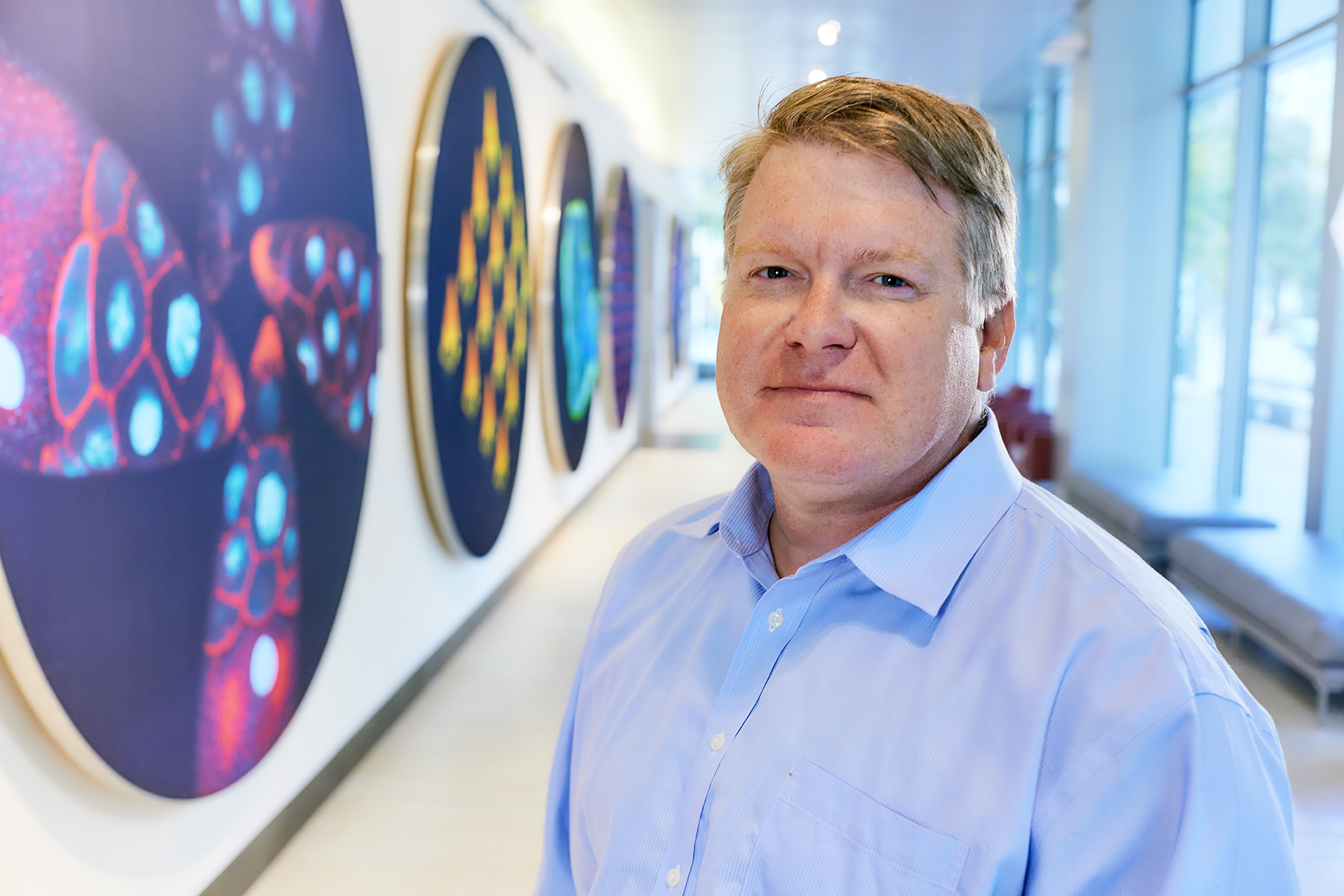
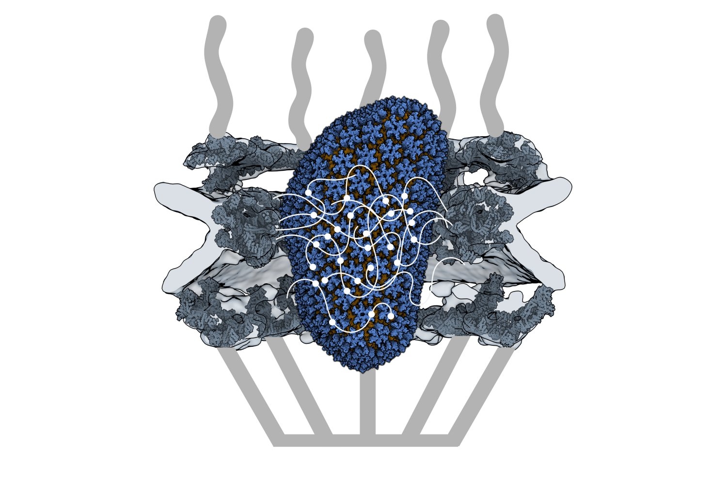
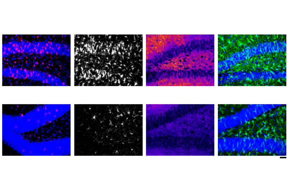
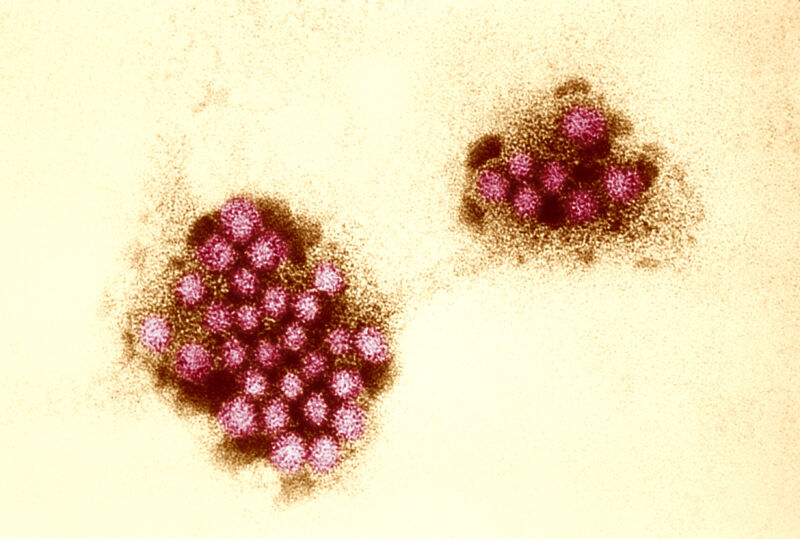


